Related Research Articles

Syphilis is a sexually transmitted infection caused by the bacterium Treponema pallidum subspecies pallidum. The signs and symptoms depend on the stage it presents: primary, secondary, latent or tertiary. The primary stage classically presents with a single chancre though there may be multiple sores. In secondary syphilis, a diffuse rash occurs, which frequently involves the palms of the hands and soles of the feet. There may also be sores in the mouth or vagina. Latent syphilis has no symptoms and can last years. In tertiary syphilis, there are gummas, neurological problems, or heart symptoms. Syphilis has been known as "the great imitator" because it may cause symptoms similar to many other diseases.
Treponema pallidum, formerly known as Spirochaeta pallida, is a microaerophilic, gram-negative, spirochaete bacterium with subspecies that cause the diseases syphilis, bejel, and yaws. It is known to be transmitted only among humans and baboons. T. pallidum can enter the host through mucosal membranes or open lesions in the skin and is primarily spread through sexual contact. It is a helically coiled microorganism usually 6–15 μm long and 0.1–0.2 μm wide. T. pallidum's lack of both a tricarboxylic acid cycle and processes for oxidative phosphorylation results in minimal metabolic activity. As a chemoorganoheterotroph, Treponema pallidum is an obligate parasite that acquires its glucose carbon source from its host. Glucose can be used not only as a primary carbon source but also in glycolytic mechanisms to generate ATP needed to power the bacterium given its minimal genome. The treponemes have cytoplasmic and outer membranes. Using light microscopy, treponemes are visible only by using dark-field illumination. T. pallidum consists of three subspecies, T. p. pallidum, T. p. endemicum, and T. p. pertenue, each of which has a distinct related disorder. The ability of T. pallidum to avoid host immune defenses has allowed for stealth pathogenicity. The unique outer membrane structure and minimal expression of surface proteins of T. pallidum has made vaccine development difficult. Treponema pallidum can be treated with high efficacy by antibiotics that inhibit bacterial cell wall synthesis such as the beta-lactam antimicrobial penicillin-G.

Yaws is a tropical infection of the skin, bones, and joints caused by the spirochete bacterium Treponema pallidum pertenue. The disease begins with a round, hard swelling of the skin, 2 to 5 cm in diameter. The center may break open and form an ulcer. This initial skin lesion typically heals after 3–6 months. After weeks to years, joints and bones may become painful, fatigue may develop, and new skin lesions may appear. The skin of the palms of the hands and the soles of the feet may become thick and break open. The bones may become misshapen. After 5 years or more, large areas of skin may die, leaving scars.
The hoof is the tip of a toe of an ungulate mammal, which is covered and strengthened with a thick and horny keratin covering. Artiodactyls are even-toed ungulates, species whose feet have an even number of digits; the ruminants with two digits are the most numerous, e.g. giraffe, deer, bison, cattle, goat, pigs, and sheep. The feet of perissodactyl mammals have an odd number of toes, e.g. the horse, the rhinoceros, and the tapir. Although hooves are limb structures primarily found in placental mammals, hadrosaurs such as Edmontosaurus possessed hoofed forelimbs. The marsupial Chaeropus also had hooves.
Skin disorders are among the most common health problems in dogs, and have many causes. The condition of a dog's skin and coat is also an important indicator of its general health. Skin disorders of dogs vary from acute, self-limiting problems to chronic or long-lasting problems requiring life-time treatment. Skin disorders may be primary or secondary in nature, making diagnosis complicated.

Bejel, or endemic syphilis, is a chronic skin and tissue disease caused by infection by the endemicum subspecies of the spirochete Treponema pallidum. Bejel is one of the "endemic treponematoses", a group that also includes yaws and pinta. Typically, endemic trepanematoses begin with localized lesions on the skin or mucous membranes. Pinta is limited to affecting the skin, whereas bejel and yaws are considered to be invasive because they can also cause disease in bone and other internal tissues.

A Jarisch–Herxheimer reaction is a sudden and typically transient reaction that may occur within 24 hours of being administered antibiotics for an infection by a spirochete, including syphilis, leptospirosis, Lyme disease, and relapsing fever. Signs and symptoms include fever, chills, shivers, feeling sick, headache, fast heart beat, low blood pressure, breathing fast, flushing of skin, muscle aches, and worsening of skin lesions. It may sometimes be mistaken as an allergy to the antibiotic.
The fluorescent treponemal antibody absorption (FTA-ABS) test is a diagnostic test for syphilis. Using antibodies specific for the Treponema pallidum species, such tests would be assumed to be more specific than non-treponemal testing such as VDRL but have been shown repeatedly to be sensitive but not specific for the diagnosis of neurosyphilis in cerebrospinal fluid (CSF). In addition, FTA-ABS turns positive earlier and remains positive longer than VDRL. Other treponemes, such as T. pertenue, may also produce a positive FTA-ABS. The ABS suffix refers particularly to a processing step used to remove nonspecific antispirochetal antibodies present in normal serum.

Bovine papular stomatitis is a zoonotic farmyard pox caused by Bovine papular stomatitis virus (BPSV), which can spread from infected cattle to cause disease in milkers, farmers and veterinarians. Generally there are usually one or multiple skin lesions, typically on the hands or forearm. The disease is generally mild.
Fusobacterium necrophorum is a species of bacteria responsible for Lemierre's syndrome. It has also been known to cause sinusitis, mastoiditis, and odontogenic infections.
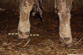
Foot rot, also known as foul-in-the-foot, interdigital necrobacillosis or infectious pododermatitis, is a hoof infection commonly found in sheep, goats, and cattle. As the name suggests, it rots away the foot of the animal, more specifically the area between the two toes of the affected animal. It is extremely painful and contagious. It can be treated with a series of medications, but if not treated, the whole herd can become infected. The cause of the infection in cattle is two species of anaerobic bacteria, Fusobacterium necrophorum and Bacteroides melaninogenicus. Both bacteria are common to the environment in which cattle live, and Fusobacterium is present in the rumen and fecal matter of the cattle. In sheep, F. necrophorum first invades the interdigital skin following damage to the skin, and causes interdigital lesions and slight inflammation. The second stage of the disease is marked by the invasion of the foot by the foot rot bacterium Dichelobacter nodosus, a Gram-negative anaerobe. Usually, an injury to the skin between the hooves allows the bacteria to infect the animal. Another cause of foot rot may be high temperatures or humidity, causing the skin between the hooves to crack and let the bacteria infect the foot. This is one of the reasons foot rot is such a major problem in the summer. Foot rot is easily identifiable by its appearance and foul odor. Treatment is usually with an antibiotic medication, and preventing injury to the feet is the best way to prevent foot rot.

Cefquinome is a fourth-generation cephalosporin with pharmacological and antibacterial properties valuable in the treatment of coliform mastitis and other infections. It is only used in veterinary applications.
Dichelobacter nodosus, formerly Bacteroides nodosus, is a Gram-negative, obligate anaerobe of the family Cardiobacteriaceae. It has polar fimbriae and is the causative agent of ovine foot rot as well as interdigital dermatitis. It is the lone species in the genus Dichelobacter.
Treponema denticola is a Gram-negative, obligate anaerobic, motile and highly proteolytic spirochete bacterium. It is one of four species of oral spirochetes to be reliably cultured, the others being Treponema pectinovorum, Treponema socranskii and Treponema vincentii. T. denticola dwells in a complex and diverse microbial community within the oral cavity and is highly specialized to survive in this environment. T. denticola is associated with the incidence and severity of human periodontal disease. Treponema denticola is one of three bacteria that form the Red Complex, the other two being Porphyromonas gingivalis and Tannerella forsythia. Together they form the major virulent pathogens that cause chronic periodontitis. Having elevated T. denticola levels in the mouth is considered one of the main etiological agents of periodontitis. T. denticola is related to the syphilis-causing obligate human pathogen, Treponema pallidum subsp. pallidum. It has also been isolated from women with bacterial vaginosis.

Tropical ulcer, more commonly known as jungle rot, is a chronic ulcerative skin lesion thought to be caused by polymicrobial infection with a variety of microorganisms, including mycobacteria. It is common in tropical climates.

Streptococcus canis is a group G beta-hemolytic species of Streptococcus. It was first isolated in dogs, giving the bacterium its name. These bacteria are characteristically different from Streptococcus dysgalactiae, which is a human-specific group G species that has a different phenotypic chemical composition. S. canis is important to the skin and mucosal health of cats and dogs, but under certain circumstances, these bacteria can cause opportunistic infections. These infections were known to afflict dogs and cats prior to the formal description of the species in Devriese et al., 1986. However, additional studies revealed cases of infection in other mammal species, including cattle and even humans. Instances of mortality from S. canis in humans are very low with only a few reported cases, while actual instances of infection may be underreported due to mischaracterizations of the bacteria as S. dysgalactiae. This species, in general, is highly susceptible to antibiotics, and plans to develop a vaccine to prevent human infections are currently being considered.
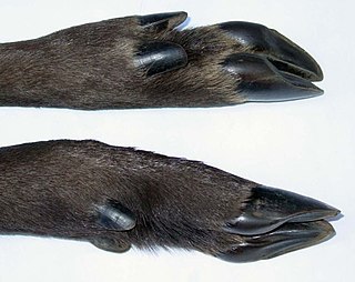
Interdigital dermatitis in cattle is caused by the anaerobic bacterium Dichelobacter nodosus. This is also the agent of footrot in sheep, but strains appear to be different and there is no cross-infection.

Staphylococcus hyicus is a Gram-positive, facultatively anaerobic bacterium in the genus Staphylococcus. It consists of clustered cocci and forms white circular colonies when grown on blood agar. S. hyicus is a known animal pathogen. It causes disease in poultry, cattle, horses, and pigs. Most notably, it is the agent that causes porcine exudative epidermitis, also known as greasy pig disease, in piglets. S. hyicus is generally considered to not be zoonotic, however it has been shown to be able to cause bacteremia and sepsis in humans.

Meningeal syphilis is a chronic form of syphilis infection that affects the central nervous system. Treponema pallidum, a spirochate bacterium, is the main cause of syphilis, which spreads drastically throughout the body and can infect all its systems if not treated appropriately. Treponema pallidum is the main cause of the onset of meningeal syphilis and other treponemal diseases, and it consists of a cytoplasmic and outer membrane that can cause a diverse array of diseases in the central nervous system and brain.
Autogenous vaccines, also called autologous vaccines, autovaccines, “self” or custom vaccines, are vaccines that are prepared by isolation and destruction of microorganisms in infected individuals and used to provide immunity to the same individual.
References
- ↑ "Dörte Döpfer on Digital Dermatitis". Archived from the original on 2016-11-26. Retrieved 2014-11-17.[ self-published source? ]
- ↑ Campbell, John (7 August 2014). "Digital dermatitis emerges in beef cattle". The Western Producer.
- ↑ Wilson-Welder, Jennifer; Alt, David; Nally, Jarlath (11 November 2015). "Digital Dermatitis in Cattle: Current Bacterial and Immunological Findings". Animals. 5 (4): 1114–1135. doi: 10.3390/ani5040400 . PMC 4693204 . PMID 26569318.
- ↑ Nally, Jarlath; Wilson-Welder, Jennifer; Alt, David (May 2015). "The etiology of digital dermatitis in ruminants: recent perspectives". Veterinary Medicine: Research and Reports. 6: 155–164. doi: 10.2147/VMRR.S62072 . PMC 6070020 . PMID 30101102.
- ↑ Döpfer, Dörte (2009). The dynamics of digital dermatitis in dairy cattle and the manageable state of disease (PDF). Proceedings of the CanWest Veterinary Conference.
- 1 2 3 Interdigital Dermatitis - Cattle [ unreliable source? ] reviewed and published by WikiVet, accessed 11 October 2011.
- ↑ Hawkins, Nik (8 November 2013). "Vet med scientists find better, safer treatments for hoof disease in cattle". University Wisconsin Madison News.
- ↑ Bjurstrom, Aerica (Spring 2016). "Walking Strong: A Fact Sheet on Dairy Hoof Health" (PDF). University Wisconsin Extension.
- ↑ "Digital Dermatitis". Archived from the original on 2014-10-23. Retrieved 2014-10-23.[ unreliable source? ]