
In archaeology, a lithic flake is a "portion of rock removed from an objective piece by percussion or pressure," and may also be referred to as simply a flake, or collectively as debitage. The objective piece, or the rock being reduced by the removal of flakes, is known as a core. Once the proper tool stone has been selected, a percussor or pressure flaker is used to direct a sharp blow, or apply sufficient force, respectively, to the surface of the stone, often on the edge of the piece. The energy of this blow propagates through the material, often producing a Hertzian cone of force which causes the rock to fracture in a controllable fashion. Since cores are often struck on an edge with a suitable angle (<90°) for flake propagation, the result is that only a portion of the Hertzian cone is created. The process continues as the flintknapper detaches the desired number of flakes from the core, which is marked with the negative scars of these removals. The surface area of the core which received the blows necessary for detaching the flakes is referred to as the striking platform.

Standard anatomical terms of location are used to describe unambiguously the anatomy of animals, including humans. The terms, typically derived from Latin or Greek roots, describe something in its standard anatomical position. This position provides a definition of what is at the front ("anterior"), behind ("posterior") and so on. As part of defining and describing terms, the body is described through the use of anatomical planes and anatomical axes.

The ulna or ulnar bone is a long bone in the forearm stretching from the elbow to the wrist. It is on the same side of the forearm as the little finger, running parallel to the radius, the forearm's other long bone. Longer and thinner than the radius, the ulna is considered to be the smaller long bone of the lower arm. The corresponding bone in the lower leg is the fibula.

In human anatomy, the wrist is variously defined as (1) the carpus or carpal bones, the complex of eight bones forming the proximal skeletal segment of the hand; (2) the wrist joint or radiocarpal joint, the joint between the radius and the carpus and; (3) the anatomical region surrounding the carpus including the distal parts of the bones of the forearm and the proximal parts of the metacarpus or five metacarpal bones and the series of joints between these bones, thus referred to as wrist joints. This region also includes the carpal tunnel, the anatomical snuff box, bracelet lines, the flexor retinaculum, and the extensor retinaculum.

The trapezoid bone is a carpal bone in tetrapods, including humans. It is the smallest bone in the distal row of carpal bones that give structure to the palm of the hand. It may be known by its wedge-shaped form, the broad end of the wedge constituting the dorsal, the narrow end the palmar surface; and by its having four articular facets touching each other, and separated by sharp edges. It is homologous with the "second distal carpal" of reptiles and amphibians.
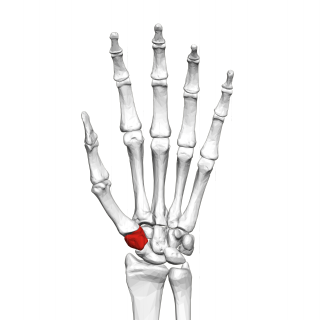
The trapezium bone is a carpal bone in the hand. It forms the radial border of the carpal tunnel.

In human anatomy, the metacarpal bones or metacarpus, also known as the "palm bones", are the appendicular bones that form the intermediate part of the hand between the phalanges (fingers) and the carpal bones, which articulate with the forearm. The metacarpal bones are homologous to the metatarsal bones in the foot.

The lunate bone is a carpal bone in the human hand. It is distinguished by its deep concavity and crescentic outline. It is situated in the center of the proximal row carpal bones, which lie between the ulna and radius and the hand. The lunate carpal bone is situated between the lateral scaphoid bone and medial triquetral bone.

The cerebrum, telencephalon or endbrain is the largest part of the brain, containing the cerebral cortex as well as several subcortical structures, including the hippocampus, basal ganglia, and olfactory bulb. In the human brain, the cerebrum is the uppermost region of the central nervous system. The cerebrum develops prenatally from the forebrain (prosencephalon). In mammals, the dorsal telencephalon, or pallium, develops into the cerebral cortex, and the ventral telencephalon, or subpallium, becomes the basal ganglia. The cerebrum is also divided into approximately symmetric left and right cerebral hemispheres.

The radius or radial bone is one of the two large bones of the forearm, the other being the ulna. It extends from the lateral side of the elbow to the thumb side of the wrist and runs parallel to the ulna. The ulna is longer than the radius, but the radius is thicker. The radius is a long bone, prism-shaped and slightly curved longitudinally.

The dorsal column–medial lemniscus pathway (DCML) (also known as the posterior column-medial lemniscus pathway is the major sensory pathway of the central nervous system that conveys sensations of fine touch, vibration, two-point discrimination, and proprioception from the skin and joints. It transmits this information to the somatosensory cortex of the postcentral gyrus in the parietal lobe of the brain. The pathway receives information from sensory receptors throughout the body, and carries this in the gracile fasciculus and the cuneate fasciculus, tracts that make up the white matter dorsal columns of the spinal cord. At the level of the medulla oblongata, the fibers of the tracts decussate and are continued in the medial lemniscus, on to the thalamus and relayed from there through the internal capsule and transmitted to the somatosensory cortex. The name dorsal-column medial lemniscus comes from the two structures that carry the sensory information: the dorsal columns of the spinal cord, and the medial lemniscus in the brainstem.

The triquetral bone is located in the wrist on the medial side of the proximal row of the carpus between the lunate and pisiform bones. It is on the ulnar side of the hand, but does not directly articulate with the ulna. Instead, it is connected to and articulates with the ulna through the Triangular fibrocartilage disc and ligament, which forms part of the ulnocarpal joint capsule. It connects with the pisiform, hamate, and lunate bones. It is the 2nd most commonly fractured carpal bone.

The mantle is a significant part of the anatomy of molluscs: it is the dorsal body wall which covers the visceral mass and usually protrudes in the form of flaps well beyond the visceral mass itself.
This glossary of ichthyology is a list of definitions of terms and concepts used in ichthyology, the study of fishes.
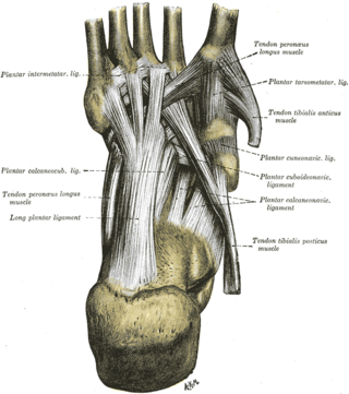
The intermetatarsal joints are the articulations between the base of metatarsal bones.

The intercarpal joints can be subdivided into three sets of joints : Those of the proximal row of carpal bones, those of the distal row of carpal bones, and those of the two rows with each other.

Gobiosuchidae is a family of Cretaceous crocodyliforms known from Mongolia and Spain.

The Panay shrew is a species of shrew from the Philippines.

Khunnuchelys was a genus of trionychine turtle from the Late Cretaceous of Asia. Three species are known, K. erinhotensis, the type species, K. kizylkumensis, and K. lophorhothon. K. erinhotensis is known from the Iren Dabasu Formation in China from the late Turonian until the middle Campanian. K. kizylkumensis is known from the late Turonian Bissekty Formation of Uzbekistan. The third species, described in 2013 by Danilov et al., is known from the early to middle Campanian aged Bostobe Formation of Kazakhstan.

Arisierpeton is an extinct genus of synapsids from the Early Permian Garber Formation of Richards Spur, Oklahoma. It contains a single species, Arisierpeton simplex.