
The carpal bones are the eight small bones that make up the wrist that connects the hand to the forearm. The term "carpus" is derived from the Latin carpus and the Greek καρπός (karpós), meaning "wrist". In human anatomy, the main role of the wrist is to facilitate effective positioning of the hand and powerful use of the extensors and flexors of the forearm, and the mobility of individual carpal bones increase the freedom of movements at the wrist.

In human anatomy, the wrist is variously defined as (1) the carpus or carpal bones, the complex of eight bones forming the proximal skeletal segment of the hand; (2) the wrist joint or radiocarpal joint, the joint between the radius and the carpus and; (3) the anatomical region surrounding the carpus including the distal parts of the bones of the forearm and the proximal parts of the metacarpus or five metacarpal bones and the series of joints between these bones, thus referred to as wrist joints. This region also includes the carpal tunnel, the anatomical snuff box, bracelet lines, the flexor retinaculum, and the extensor retinaculum.
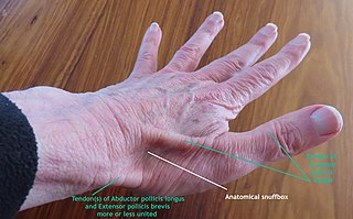
The anatomical snuff box or snuffbox or foveola radialis is a triangular deepening on the radial, dorsal aspect of the hand—at the level of the carpal bones, specifically, the scaphoid and trapezium bones forming the floor. The name originates from the use of this surface for placing and then sniffing powdered tobacco, or "snuff." It is sometimes referred to by its French name tabatière.
The forearm is the region of the upper limb between the elbow and the wrist. The term forearm is used in anatomy to distinguish it from the arm, a word which is used to describe the entire appendage of the upper limb, but which in anatomy, technically, means only the region of the upper arm, whereas the lower "arm" is called the forearm. It is homologous to the region of the leg that lies between the knee and the ankle joints, the crus.

The scaphoid bone is one of the carpal bones of the wrist. It is situated between the hand and forearm on the thumb side of the wrist. It forms the radial border of the carpal tunnel. The scaphoid bone is the largest bone of the proximal row of wrist bones, its long axis being from above downward, lateralward, and forward. It is approximately the size and shape of a medium cashew nut.

The capitate bone is a bone in the human wrist found in the center of the carpal bone region, located at the distal end of the radius and ulna bones. It articulates with the third metacarpal bone and forms the third carpometacarpal joint. The capitate bone is the largest of the carpal bones in the human hand. It presents, above, a rounded portion or head, which is received into the concavity formed by the scaphoid and lunate bones; a constricted portion or neck; and below this, the body. The bone is also found in many other mammals, and is homologous with the "third distal carpal" of reptiles and amphibians.

The lunate bone is a carpal bone in the human hand. It is distinguished by its deep concavity and crescentic outline. It is situated in the center of the proximal row carpal bones, which lie between the ulna and radius and the hand. The lunate carpal bone is situated between the lateral scaphoid bone and medial triquetral bone.

A Colles' fracture is a type of fracture of the distal forearm in which the broken end of the radius is bent backwards. Symptoms may include pain, swelling, deformity, and bruising. Complications may include damage to the median nerve.
A distal radius fracture, also known as wrist fracture, is a break of the part of the radius bone which is close to the wrist. Symptoms include pain, bruising, and rapid-onset swelling. The ulna bone may also be broken.

A Smith's fracture, is a fracture of the distal radius.

Madelung's deformity is usually characterized by malformed wrists and wrist bones and is often associated with Léri-Weill dyschondrosteosis. It can be bilateral or just in the one wrist. It has only been recognized within the past hundred years. Named after Otto Wilhelm Madelung (1846–1926), a German surgeon, who described it in detail, it was noted by others. Guillaume Dupuytren mentioned it in 1834, Auguste Nélaton in 1847, and Joseph-François Malgaigne in 1855.

The radial styloid process is a projection of bone on the lateral surface of the distal radius bone.

The triangular fibrocartilage complex (TFCC) is formed by the triangular fibrocartilage discus (TFC), the radioulnar ligaments (RULs) and the ulnocarpal ligaments (UCLs).

A scaphoid fracture is a break of the scaphoid bone in the wrist. Symptoms generally includes pain at the base of the thumb which is worse with use of the hand. The anatomic snuffbox is generally tender and swelling may occur. Complications may include nonunion of the fracture, avascular necrosis of the proximal part of the bone, and arthritis.

The scapholunate ligament is a ligament of the wrist.

A hand is a prehensile, multi-fingered appendage located at the end of the forearm or forelimb of primates such as humans, chimpanzees, monkeys, and lemurs. A few other vertebrates such as the koala are often described as having "hands" instead of paws on their front limbs. The raccoon is usually described as having "hands" though opposable thumbs are lacking.
Watson's test, also known as the scaphoid shift test, is a diagnostic test for instability between the scaphoid and lunate bones of the wrist.

Wrist osteoarthritis is a group of mechanical abnormalities resulting in joint destruction, which can occur in the wrist. These abnormalities include degeneration of cartilage and hypertrophic bone changes, which can lead to pain, swelling and loss of function. Osteoarthritis of the wrist is one of the most common conditions seen by hand surgeons.

Wrist arthroscopy can be used to look inside the joint of the wrist. It is a minimally invasive technique which can be utilized for diagnostic purposes as well as for therapeutic interventions. Wrist arthroscopy has been used for diagnostic purposes since it was first introduced in 1979. However, it only became accepted as diagnostic tool around the mid-1980s. At that time, arthroscopy of the wrist was an innovative technique to determine whether a problem could be found in the wrist. A few years later, wrist arthroscopy could also be used as a therapeutic tool.

Lister's tubercle or dorsal tubercle of radius is a bony prominence located at the distal end of the radius. It is palpable on the dorsum of the wrist.
