Related Research Articles
Medical physics deals with the application of the concepts and methods of physics to the prevention, diagnosis and treatment of human diseases with a specific goal of improving human health and well-being. Since 2008, medical physics has been included as a health profession according to International Standard Classification of Occupation of the International Labour Organization.

Tomography is imaging by sections or sectioning that uses any kind of penetrating wave. The method is used in radiology, archaeology, biology, atmospheric science, geophysics, oceanography, plasma physics, materials science, cosmochemistry, astrophysics, quantum information, and other areas of science. The word tomography is derived from Ancient Greek τόμος tomos, "slice, section" and γράφω graphō, "to write" or, in this context as well, "to describe." A device used in tomography is called a tomograph, while the image produced is a tomogram.

Electrical impedance tomography (EIT) is a noninvasive type of medical imaging in which the electrical conductivity, permittivity, and impedance of a part of the body is inferred from surface electrode measurements and used to form a tomographic image of that part. Electrical conductivity varies considerably among various biological tissues or the movement of fluids and gases within tissues. The majority of EIT systems apply small alternating currents at a single frequency, however, some EIT systems use multiple frequencies to better differentiate between normal and suspected abnormal tissue within the same organ.
Medical optical imaging is the use of light as an investigational imaging technique for medical applications, pioneered by American Physical Chemist Britton Chance. Examples include optical microscopy, spectroscopy, endoscopy, scanning laser ophthalmoscopy, laser Doppler imaging, and optical coherence tomography. Because light is an electromagnetic wave, similar phenomena occur in X-rays, microwaves, and radio waves.

Functional near-infrared spectroscopy (fNIRS) is an optical brain monitoring technique which uses near-infrared spectroscopy for the purpose of functional neuroimaging. Using fNIRS, brain activity is measured by using near-infrared light to estimate cortical hemodynamic activity which occur in response to neural activity. Alongside EEG, fNIRS is one of the most common non-invasive neuroimaging techniques which can be used in portable contexts. The signal is often compared with the BOLD signal measured by fMRI and is capable of measuring changes both in oxy- and deoxyhemoglobin concentration, but can only measure from regions near the cortical surface. fNIRS may also be referred to as Optical Topography (OT) and is sometimes referred to simply as NIRS.

Neuroimaging is the use of quantitative (computational) techniques to study the structure and function of the central nervous system, developed as an objective way of scientifically studying the healthy human brain in a non-invasive manner. Increasingly it is also being used for quantitative research studies of brain disease and psychiatric illness. Neuroimaging is highly multidisciplinary involving neuroscience, computer science, psychology and statistics, and is not a medical specialty. Neuroimaging is sometimes confused with neuroradiology.

Electrical resistivity tomography (ERT) or electrical resistivity imaging (ERI) is a geophysical technique for imaging sub-surface structures from electrical resistivity measurements made at the surface, or by electrodes in one or more boreholes. If the electrodes are suspended in the boreholes, deeper sections can be investigated. It is closely related to the medical imaging technique electrical impedance tomography (EIT), and mathematically is the same inverse problem. In contrast to medical EIT, however, ERT is essentially a direct current method. A related geophysical method, induced polarization, measures the transient response and aims to determine the subsurface chargeability properties.

Optical tomography is a form of computed tomography that creates a digital volumetric model of an object by reconstructing images made from light transmitted and scattered through an object. Optical tomography is used mostly in medical imaging research. Optical tomography in industry is used as a sensor of thickness and internal structure of semiconductors.

Electrical capacitance tomography (ECT) is a method for determination of the dielectric permittivity distribution in the interior of an object from external capacitance measurements. It is a close relative of electrical impedance tomography and is proposed as a method for industrial process monitoring.
Process tomography consists of tomographic imaging of systems, such as process pipes in industry. In tomography the 3D distribution of some physical quantity in the object is determined. There is a widespread need to get tomographic information about process. This information can be used, for example, in the design and control of processes.
Focused Impedance Measurement (FIM) is a recent technique for quantifying the electrical resistance in tissues of the human body with improved zone localization compared to conventional methods. This method was proposed and developed by Department of Biomedical Physics and Technology of University of Dhaka under the supervision of Prof. Khondkar Siddique-e-Rabbani; who first introduced the idea. FIM can be considered a bridge between Four Electrode Impedance Measurement (FEIM) and Electrical impedance Tomography (EIT), and provides a middle ground in terms of simplicity and accuracy.
Lassi Päivärinta is a Finnish mathematician, one-time professor of applied mathematics at the department of mathematics and statistics at the University of Helsinki. Päivärinta's research is mostly in the fields of inverse problems and partial differential equations.
Electrical cardiometry is a method based on the model of Electrical Velocimetry, and non-invasively measures stroke volume (SV), cardiac output (CO), and other hemodynamic parameters through the use of 4 surface ECG electrodes. Electrical cardiometry is a method trademarked by Cardiotronic, Inc., and is U.S. FDA approved for use on adults, children, and neonates.
Boundary estimation in EIT is the term used in the field of electrical impedance tomography, if the inverse problem is the estimation of boundary instead of the conductivity distribution inside an object domain.

Diffuse optical imaging (DOI) is a method of imaging using near-infrared spectroscopy (NIRS) or fluorescence-based methods. When used to create 3D volumetric models of the imaged material DOI is referred to as diffuse optical tomography, whereas 2D imaging methods are classified as diffuse optical imaging.

Industrial Tomography Systems plc, occasionally abbreviated to ITOMS or simply ITS, is a manufacturer of process visualization systems based upon the principles of tomography. Headquartered in Manchester, UK, the company provides instrumentation to a variety of organisations across a range of sectors; including oil refining, chemical manufacturing, nuclear engineering, dairy manufacturing, and research/academia.

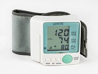
A medical procedure is defined as non-invasive when no break in the skin is created and there is no contact with the mucosa, or skin break, or internal body cavity beyond a natural or artificial body orifice. For example, deep palpation and percussion are non-invasive but a rectal examination is invasive. Likewise, examination of the ear-drum or inside the nose or a wound dressing change all fall outside the definition of non-invasive procedure. There are many non-invasive procedures, ranging from simple observation, to specialised forms of surgery, such as radiosurgery. Extracorporeal shock wave lithotripsy is a non-invasive treatment of stones in the kidney, gallbladder or liver, using an acoustic pulse. For centuries, physicians have employed many simple non-invasive methods based on physical parameters in order to assess body function in health and disease, such as pulse-taking, the auscultation of heart sounds and lung sounds, temperature examination, respiratory examination, peripheral vascular examination, oral examination, abdominal examination, external percussion and palpation, blood pressure measurement, change in body volumes, audiometry, eye examination, and many others.
Impedance microbiology is a microbiological technique used to measure the microbial number density of a sample by monitoring the electrical parameters of the growth medium. The ability of microbial metabolism to change the electrical conductivity of the growth medium was discovered by Stewart and further studied by other scientists such as Oker-Blom, Parson and Allison in the first half of 20th century. However, it was only in the late 1970s that, thanks to computer-controlled systems used to monitor impedance, the technique showed its full potential, as discussed in the works of Fistenberg-Eden & Eden, Ur & Brown and Cady.
Electrical capacitance volume tomography (ECVT) is a non-invasive 3D imaging technology applied primarily to multiphase flows. It was first introduced by W. Warsito, Q. Marashdeh, and L.-S. Fan as an extension of the conventional electrical capacitance tomography (ECT). In conventional ECT, sensor plates are distributed around a surface of interest. Measured capacitance between plate combinations is used to reconstruct 2D images (tomograms) of material distribution. In ECT, the fringing field from the edges of the plates is viewed as a source of distortion to the final reconstructed image and is thus mitigated by guard electrodes. ECVT exploits this fringing field and expands it through 3D sensor designs that deliberately establish an electric field variation in all three dimensions. The image reconstruction algorithms are similar in nature to ECT; nevertheless, the reconstruction problem in ECVT is more complicated. The sensitivity matrix of an ECVT sensor is more ill-conditioned and the overall reconstruction problem is more ill-posed compared to ECT. The ECVT approach to sensor design allows direct 3D imaging of the outrounded geometry. This is different than 3D-ECT that relies on stacking images from individual ECT sensors. 3D-ECT can also be accomplished by stacking frames from a sequence of time intervals of ECT measurements. Because the ECT sensor plates are required to have lengths on the order of the domain cross-section, 3D-ECT does not provide the required resolution in the axial dimension. ECVT solves this problem by going directly to the image reconstruction and avoiding the stacking approach. This is accomplished by using a sensor that is inherently three-dimensional.
Diffuse optical mammography, or simply optical mammography, is an emerging imaging technique that enables the investigation of the breast composition through spectral analysis. It combines in a single non-invasive tool the capability to implement breast cancer risk assessment, lesion characterization, therapy monitoring and prediction of therapy outcome. It is an application of diffuse optics, which studies light propagation in strongly diffusive media, such as biological tissues, working in the red and near-infrared spectral range, between 600 and 1100 nm.
References
- ↑ W R B Lionheart, S R Arridge, M Schweiger, M Vauhkonen and J P Kaipio, Electrical Impedance and Diffuse Optical Tomography Reconstruction Software, Proceedings of the 1st World Congress on Industrial Process Tomography, pp 474–477, Buxton, Derbyshire, 1999
- ↑ Vauhkonen, P. J., Vauhkonen, M., Savolainen, T., & Kaipio, J. P. (1999). Three-dimensional electrical impedance tomography based on the complete electrode model. Biomedical Engineering, IEEE Transactions on, 46(9), 1150–1160.
- ↑ Polydorides N, Lionheart WRB, A Matlab toolkit for three-dimensional electrical impedance tomography: a contribution to the Electrical Impedance and Diffuse Optical Reconstruction Software project, Meas. Sci. Technol. 13 (December 2002) 1871–1883
- ↑ A Adler and W R B Lionheart, Uses and abuses of EIDORS: An extensible software base for EIT, Physiol Meas 27, S25–S42, 2006.
- ↑ A. Biguri; B. Grychtol; A. Adler; M. Soleimani (2015). "Tracking boundary movement and exterior shape modelling in lung EIT imaging" (PDF). Physiological Measurement. 36 (6): 1119–35. Bibcode:2015PhyM...36.1119B. doi:10.1088/0967-3334/36/6/1119. PMID 26007150. S2CID 30064176.
- ↑ Martin Proença et al., Influence of heart motion on cardiac output estimation by means of electrical impedance tomography: a case study Physiological Measurement 2015 36 1075
- ↑ Kirill Y. Aristovich et al Imaging fast electrical activity in the brain with electrical impedance tomography NeuroImage 2016 124 204
- ↑ Hun Wi et al. Real-time conductivity imaging of temperature and tissue property changes during radiofrequency ablation: An ex vivo model using weighted frequency difference Bioelectromagnetics 2015 36 277
- ↑ Kent Wei et al., ITS Reconstruction Tool-Suite: An inverse algorithm package for industrial process tomography Flow Measurement and Instrumentation 2015 46 292
- ↑ Suze-Anne Korteland and Timo Heimovaara, Quantitative inverse modelling of a cylindrical object in the laboratory using ERT: An error analysis Journal of Applied Geophysics 2015
- ↑ Gerard J. Gallo and Erik T. Thostenson Spatial damage detection in electrically anisotropic fiber-reinforced composites using carbon nanotube networks Composite Structures 2015