Related Research Articles

Magnetoencephalography (MEG) is a functional neuroimaging technique for mapping brain activity by recording magnetic fields produced by electrical currents occurring naturally in the brain, using very sensitive magnetometers. Arrays of SQUIDs are currently the most common magnetometer, while the SERF magnetometer is being investigated for future machines. Applications of MEG include basic research into perceptual and cognitive brain processes, localizing regions affected by pathology before surgical removal, determining the function of various parts of the brain, and neurofeedback. This can be applied in a clinical setting to find locations of abnormalities as well as in an experimental setting to simply measure brain activity.

Functional neuroimaging is the use of neuroimaging technology to measure an aspect of brain function, often with a view to understanding the relationship between activity in certain brain areas and specific mental functions. It is primarily used as a research tool in cognitive neuroscience, cognitive psychology, neuropsychology, and social neuroscience.
Neuroimaging is a medical technique that allows doctors and researchers to take pictures of the inner workings of the body or brain of a patient. It can show areas with heightened activity, areas with high or low blood flow, the structure of the patients brain/body, as well as certain abnormalities. Neuroimaging is most often used to find the specific location of certain diseases or birth defects such as tumors, cancers, or clogged arteries. Neuroimaging first came about as a medical technique in the 1880s with the invention of the human circulation balance and has since lead to other inventions such as the x-ray, air ventriculography, cerebral angiography, PET/SPECT scans, magnetoencephalography, and xenon CT scanning.

The superior temporal gyrus (STG) is one of three gyri in the temporal lobe of the human brain, which is located laterally to the head, situated somewhat above the external ear.
Functional integration is the study of how brain regions work together to process information and effect responses. Though functional integration frequently relies on anatomic knowledge of the connections between brain areas, the emphasis is on how large clusters of neurons – numbering in the thousands or millions – fire together under various stimuli. The large datasets required for such a whole-scale picture of brain function have motivated the development of several novel and general methods for the statistical analysis of interdependence, such as dynamic causal modelling and statistical linear parametric mapping. These datasets are typically gathered in human subjects by non-invasive methods such as EEG/MEG, fMRI, or PET. The results can be of clinical value by helping to identify the regions responsible for psychiatric disorders, as well as to assess how different activities or lifestyles affect the functioning of the brain.

Neuroimaging is the use of quantitative (computational) techniques to study the structure and function of the central nervous system, developed as an objective way of scientifically studying the healthy human brain in a non-invasive manner. Increasingly it is also being used for quantitative research studies of brain disease and psychiatric illness. Neuroimaging is highly multidisciplinary involving neuroscience, computer science, psychology and statistics, and is not a medical specialty. Neuroimaging is sometimes confused with neuroradiology.
The Cognition and Brain Sciences Unit is a branch of the UK Medical Research Council, based in Cambridge, England. The CBSU is a centre for cognitive neuroscience, with a mission to improve human health by understanding and enhancing cognition and behaviour in health, disease and disorder. It is one of the largest and most long-lasting contributors to the development of psychological theory and practice.
Thalamocortical dysrhythmia (TCD) is a theoretical framework in which neuroscientists try to explain the positive and negative symptoms induced by neuropsychiatric disorders like Parkinson's Disease, neurogenic pain, tinnitus, visual snow syndrome, schizophrenia, obsessive–compulsive disorder, depressive disorder and epilepsy. In TCD, normal thalamocortical resonance is disrupted by changes in the behaviour of neurons in the thalamus.
TCD can be treated with neurosurgical methods like the central lateral thalamotomy, which due to its invasiveness is only used on patients that have proven resistant to conventional therapies.
A medical procedure is a course of action intended to achieve a result in the delivery of healthcare.
Synthetic-aperture magnetometry (SAM) is a method for analysis of data obtained from magnetoencephalography (MEG) and electroencephalography (EEG). SAM is a nonlinear beamforming approach which can be thought of as a spatial filter.
The Athinoula A. Martinos Center for Biomedical Imaging, usually referred to as just the "Martinos Center," is a major hub of biomedical imaging technology development and translational research. The Center is part of the Department of Radiology at Massachusetts General Hospital and is affiliated with both Harvard University and MIT. Bruce Rosen is the Director of the Center and Monica Langone is the Administrative Director.
The following outline is provided as an overview of and topical guide to brain mapping:

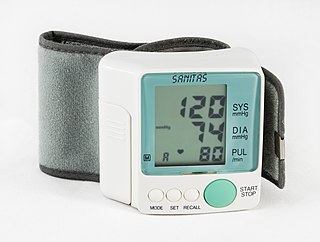
A medical procedure is defined as non-invasive when no break in the skin is created and there is no contact with the mucosa, or skin break, or internal body cavity beyond a natural or artificial body orifice. For example, deep palpation and percussion are non-invasive but a rectal examination is invasive. Likewise, examination of the ear-drum or inside the nose or a wound dressing change all fall outside the definition of non-invasive procedure. There are many non-invasive procedures, ranging from simple observation, to specialised forms of surgery, such as radiosurgery. Extracorporeal shock wave lithotripsy is a non-invasive treatment of stones in the kidney, gallbladder or liver, using an acoustic pulse. For centuries, physicians have employed many simple non-invasive methods based on physical parameters in order to assess body function in health and disease, such as pulse-taking, the auscultation of heart sounds and lung sounds, temperature examination, respiratory examination, peripheral vascular examination, oral examination, abdominal examination, external percussion and palpation, blood pressure measurement, change in body volumes, audiometry, eye examination, and many others.
Bruce Rosen is an American physicist and radiologist and a leading expert in the area of functional neuroimaging. His research for the past 30 years has focused on the development and application of physiological and functional nuclear magnetic resonance techniques, as well as new approaches to combine functional magnetic resonance imaging (fMRI) data with information from other modalities such as positron emission tomography (PET), magnetoencephalography (MEG) and noninvasive optical imaging. The techniques his group has developed to measure physiological and metabolic changes associated with brain activation and cerebrovascular insult are used by research centers and hospitals throughout the world.
The Ludwig-Boltzmann-Institute for functional Brain Topography was a research institute for the investigation of the function of brain areas. It was founded in 1993 in Vienna, Austria by Lüder Deecke. With his retirement in 2006 the institute was closed.

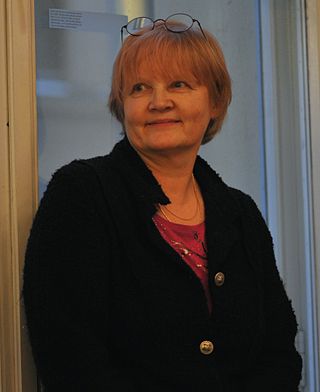
Riitta Kyllikki Hari is a Finnish neuroscientist, physician and professor at Aalto University. She has led the Brain Research Unit at the Low Temperature Laboratory since 1982. Hari was appointed as Academician of Science on 26 November 2010.

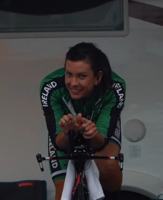
Kelly Murphy is an Irish racing cyclist, who currently rides for UCI Women's Continental Team IBCT. She was elite women's national champion in the time trial event in 2018 & 2019 and represented Ireland in this event at the European and World Championships in these same years. At the 2019 European Road Championships, Murphy finished 10th in the TT and in doing so became the first Irish woman to place within the top 10 at any international road event.
Ole Jensen is a Danish neuroscientist and professor of translational neuroscience at the School of Psychology, University of Birmingham. He is known for his research work on applying magnetoencephalography to study the functioning of human brain.
Michelle Anna Espy is an American physicist at the Los Alamos National Laboratory who studies ultra-low-field nuclear magnetic resonance magnetic resonance imaging using SQUIDs, with applications including magnetoencephalography and the detection of explosive materials and nerve agents in airline security screening. At Los Alamos, she has also worked on neutron imaging of stockpiled weapons and of the skull of the Bisti Beast, a fossil tyrannosaur.
References
- ↑ Akalin, Zeynep; Makeig, Scott. "REALISTIC MODELING OF THE HUMAN HEAD FROM AVAILABLE INFORMATION" (PDF). Retrieved 2 November 2018.