Equivalence group

Last updated

An equivalence group is a set of unspecified cells that have the same developmental potential or ability to adopt various fates. Our[ who? ] current understanding suggests that equivalence groups are limited to cells of the same ancestry, also known as sibling cells. [1] Often, cells of an equivalence group adopt different fates from one another. [2]

Contents

Equivalence groups assume various potential fates in two general, non-mutually exclusive ways. One mechanism, induction, occurs when a signal originating from outside of the equivalence group specifies a subset of the naïve cells. [2] Another mode, known as lateral inhibition, arises when a signal within an equivalence group causes one cell to adopt a dominant fate while others in the group are inhibited from doing so. [3] In many examples of equivalence groups, both induction and lateral inhibition are used to define patterns of distinct cell types.

Cells of an equivalence group that do not receive a signal adopt a default fate. Alternatively, cells that receive a signal take on different fates. [2] [4] At a certain point, the fates of cells within an equivalence group become irreversibly determined, thus they lose their multipotent potential. The following provides examples of equivalence groups studied in nematodes and ascidians.

Vulva Precursor Cell Equivalence Group

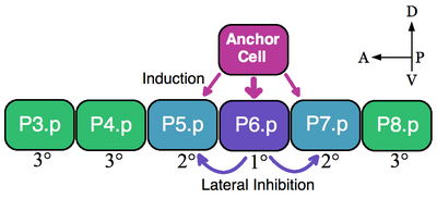
Model for the determination of the VPCs (cells P3.p-P8.p) in C. elegans. An inductive signal from the anchor cell via LIN-3 determines P6.p to adopt the 1deg. Then, the lateral signal LIN-12 causes P5.p and P5.7 to become 2deg. The other VPCs do not receive an inductive signal and are fated as 3deg. Anterior is to the left. VPCs.png
Model for the determination of the VPCs (cells P3.p-P8.p) in C. elegans. An inductive signal from the anchor cell via LIN-3 determines P6.p to adopt the 1°. Then, the lateral signal LIN-12 causes P5.p and P5.7 to become 2°. The other VPCs do not receive an inductive signal and are fated as 3°. Anterior is to the left.

Introduction

A classic example of an equivalence group is the vulva precursor cells (VPCs) of nematodes. In Caenorhabditis elegans self-fertilized eggs exit the body through the vulva. This organ develops from a subset of cell of an equivalence group consisting of six VPCs, P3.p-P8.p, which lie ventrally along the anterior-posterior axis. [5] In this example a single overlying somatic cells, the anchor cell, induces nearby VPCs to take on vulva fates 1° (P6.p) and 2° (P5.p and P7.p). VPCs that are not induced form the 3° lineage (P3.p, P4.p and P8.p), which make epidermal cells that fuse to a large syncytial epidermis (see image). [6]

The six VPCs form an equivalence group because all of the six cells are competent to take on any of the available fates (1°, 2°, and 3°) dependent on their proximity to the anchor cell. Ablation experiments indicate that all VPCs are able to adopt vulva fates. For example, if the P6.p cell that normally becomes 1° is ablated then the VPC closest to the anchor cell, either P5.p or P7.p, assumes the 1° fate. Furthermore, if all VPCs are destroyed except the most anterior P3.p cell then the anchor cell designates this cell the 1° fate. However, if the anchor cell is killed, in the absence of an inductive signal, then all of the VPCs assume the default 3° lineage. [7]

Molecular Mechanism

The anchor cell directly induces the vulva fates by secreting the epidermal growth factor (EGF)-like ligand LIN-3. The P6.p cell receives the LIN-3 signal via the receptor tyrosine kinase LET-23 (P5.p and P7.p also receive LIN-3 but to a lesser extent). Activation of LET-23 in P6.p results in the activation of LIN-12 (Notch) in P5.p and P7.p. Experimental evidence shows that LIN-12 is necessary and sufficient for the formation of the 2° fate. Through lateral inhibition LIN-12 prevents the P5.p and P7.p cells from adopting the 1° lineage. [7] Thus, in this example both inductive EGF signaling and lateral Notch activation patterns the VPC equivalence group.

Ascidian Pigment Precursor Equivalence Group

Two bilateral pigment precursor cells form an equivalence group that eventually interacts to seal mutually exclusive fates (otolith and ocellus). Shown is a timeline of the developing Halocythia roretzi embryo (anterior is to the left). Pigment precursor cells (gray circles) remain equivalent until they align after neurulation. At 21 hours post fertilization the ocellus (white circle) and otolith (black circle) are determined. Otolith Ocellus.png
Two bilateral pigment precursor cells form an equivalence group that eventually interacts to seal mutually exclusive fates (otolith and ocellus). Shown is a timeline of the developing Halocythia roretzi embryo (anterior is to the left). Pigment precursor cells (gray circles) remain equivalent until they align after neurulation. At 21 hours post fertilization the ocellus (white circle) and otolith (black circle) are determined.

Introduction

The larvae of ascidians (sea squirts) contain a pair of sensory pigment cells known as the otolith and ocellus. The otolith is used to sense gravity, whereas the ocellus responds to light. During embryogenesis the otolith and ocellus develop from two bilateral equivalent precursors. Either the left or right pigment precursor cell has equal probability of developing into the otolith or ocellus. The decision to adopt either fate is determined after neural tube closure during the early tailbud stage (see image), via a poorly defined mechanism of induction. [1]

During normal development, after neural tube closure, the pigment precursors align dorsally along the anterior-posterior axis of the neural tube. Whichever cell aligns anteriorly will become the otolith, while the posterior cell will form the ocellus. In the absence of cell-cell interactions both cells develop into ocelli, which is the default fate. [3] [8]

Experimental Methods for Studying Equivalence in Halocynthia roretzi

To elucidate whether the fates of the otolith and ocellus are determined in the early embryo or after the precursors align during neural tube closure, ablation and drug treatment techniques were used in the ascidian species Halocythia roretzi .

Cells that are labeled with fluorescein isothiocyanate-dextran (FDX) can be selectively photoablated by fluorescent excitation. [9] When one FDX labeled pigment precursor cells is photoablated during the mid-neurula stage (15 hrs) the other will almost always develop into an ocellus. However, if the ablations are performed during the late tailbud stage (22.5 hrs) then the remaining cell has an equal likelihood of becoming an otolith or ocellus. [1]

Inhibiting cell division and morphogenesis with cytochalasin B is another method used to determine when the pigment precursor equivalence group is specified. Cytochalasin treatment of early tailbud stage embryos (17 hrs), while the two bilateral cells are still separated, results in both cells becoming ocelli. When the drug was used after the two cells aligned at the dorsal midline, the anterior cell developed into the otolith and the posterior cell became the ocellus without exception. [1] Both experiments suggest that fates of the pigment precursor cells are irreversibly determined by approximately the mid-tailbud stage (21 hrs).

Other Equivalence Groups

Equivalence groups have also been described in the ganglion mother cells in grasshopper and the O/P teloblasts in the leech. [10] [11] Like other instances of equivalence groups, progeny cells are born equivalent and become specified through cell interactions. Equivalence groups are a common theme in the development of many organisms from diverse phyla.

Related Research Articles

<span class="mw-page-title-main">Gastrulation</span> Stage in embryonic development in which germ layers form

Gastrulation is the stage in the early embryonic development of most animals, during which the blastula, or in mammals the blastocyst is reorganized into a multilayered structure known as the gastrula. Before gastrulation, the embryo is a continuous epithelial sheet of cells; by the end of gastrulation, the embryo has begun differentiation to establish distinct cell lineages, set up the basic axes of the body, and internalized one or more cell types including the prospective gut.

Segmentation in biology is the division of some animal and plant body plans into a series of repetitive segments. This article focuses on the segmentation of animal body plans, specifically using the examples of the taxa Arthropoda, Chordata, and Annelida. These three groups form segments by using a "growth zone" to direct and define the segments. While all three have a generally segmented body plan and use a growth zone, they use different mechanisms for generating this patterning. Even within these groups, different organisms have different mechanisms for segmenting the body. Segmentation of the body plan is important for allowing free movement and development of certain body parts. It also allows for regeneration in specific individuals.
<span class="mw-page-title-main">Notch signaling pathway</span> Series of molecular signals

The Notch signaling pathway is a highly conserved cell signaling system present in most animals. Mammals possess four different notch receptors, referred to as NOTCH1, NOTCH2, NOTCH3, and NOTCH4. The notch receptor is a single-pass transmembrane receptor protein. It is a hetero-oligomer composed of a large extracellular portion, which associates in a calcium-dependent, non-covalent interaction with a smaller piece of the notch protein composed of a short extracellular region, a single transmembrane-pass, and a small intracellular region.

<span class="mw-page-title-main">Neural crest</span> Pluripotent embyronic cell group giving rise to diverse cell lineages

Neural crest cells are a temporary group of cells that arise from the embryonic ectoderm germ layer, and in turn give rise to a diverse cell lineage—including melanocytes, craniofacial cartilage and bone, smooth muscle, peripheral and enteric neurons and glia.

<span class="mw-page-title-main">Primitive streak</span> Structure in early amniote embryogenesis

The primitive streak is a structure that forms in the early embryo in amniotes. In amphibians the equivalent structure is the blastopore. During early embryonic development, the embryonic disc becomes oval shaped, and then pear-shaped with the broad end towards the anterior, and the narrower region projected to the posterior. The primitive streak forms a longitudinal midline structure in the narrower posterior (caudal) region of the developing embryo on its dorsal side. At first formation the primitive streak extends for half the length of the embryo. In the human embryo this appears by stage 6, about 17 days.

Compartments can be simply defined as separate, different, adjacent cell populations, which upon juxtaposition, create a lineage boundary. This boundary prevents cell movement from cells from different lineages across this barrier, restricting them to their compartment. Subdivisions are established by morphogen gradients and maintained by local cell-cell interactions, providing functional units with domains of different regulatory genes, which give rise to distinct fates. Compartment boundaries are found across species. In the hindbrain of vertebrate embryos, rhombomeres are compartments of common lineage outlined by expression of Hox genes. In invertebrates, the wing imaginal disc of Drosophila provides an excellent model for the study of compartments. Although other tissues, such as the abdomen, and even other imaginal discs are compartmentalized, much of our understanding of key concepts and molecular mechanisms involved in compartment boundaries has been derived from experimentation in the wing disc of the fruit fly.

An asymmetric cell division produces two daughter cells with different cellular fates. This is in contrast to symmetric cell divisions which give rise to daughter cells of equivalent fates. Notably, stem cells divide asymmetrically to give rise to two distinct daughter cells: one copy of the original stem cell as well as a second daughter programmed to differentiate into a non-stem cell fate.

Within the field of developmental biology, one goal is to understand how a particular cell develops into a final cell type, known as fate determination. Within an embryo, several processes play out at the cellular and tissue level to create an organism. These processes include cell proliferation, differentiation, cellular movement and programmed cell death. Each cell in an embryo receives molecular signals from neighboring cells in the form of proteins, RNAs and even surface interactions. Almost all animals undergo a similar sequence of events during very early development, a conserved process known as embryogenesis. During embryogenesis, cells exist in three germ layers, and undergo gastrulation. While embryogenesis has been studied for more than a century, it was only recently that scientists discovered that a basic set of the same proteins and mRNAs are involved in embryogenesis. Evolutionary conservation is one of the reasons that model systems such as the fly, the mouse, and other organisms are used as models to study embryogenesis and developmental biology. Studying model organisms provides information relevant to other animals, including humans. While studying the different model systems, cells fate was discovered to be determined via multiple ways, two of which are by the combination of transcription factors the cells have and by the cell-cell interaction. Cells’ fate determination mechanisms were categorized into three different types, autonomously specified cells, conditionally specified cells, or syncytial specified cells. Furthermore, the cells’ fate was determined mainly using two types of experiments, cell ablation and transplantation. The results obtained from these experiments, helped in identifying the fate of the examined cells.

In the field of developmental biology, regional differentiation is the process by which different areas are identified in the development of the early embryo. The process by which the cells become specified differs between organisms.

The scleraxis protein is a member of the basic helix-loop-helix (bHLH) superfamily of transcription factors. Currently two genes have been identified to code for identical scleraxis proteins.

<span class="mw-page-title-main">Fate mapping</span>

Fate mapping is a method used in developmental biology to study the embryonic origin of various adult tissues and structures. The "fate" of each cell or group of cells is mapped onto the embryo, showing which parts of the embryo will develop into which tissue. When carried out at single-cell resolution, this process is called cell lineage tracing. It is also used to trace the development of tumors.

Convergent extension (CE), sometimes called convergence and extension (C&E), is the process by which the tissue of an embryo is restructured to converge (narrow) along one axis and extend (elongate) along a perpendicular axis by cellular movement.

<span class="mw-page-title-main">NUMB (gene)</span> Protein-coding gene in the species Homo sapiens

Protein numb homolog is a protein that in humans is encoded by the NUMB gene. The protein encoded by this gene plays a role in the determination of cell fates during development. The encoded protein, whose degradation is induced in a proteasome-dependent manner by MDM2, is a membrane-bound protein that has been shown to associate with EPS15, LNX1, and NOTCH1. Four transcript variants encoding different isoforms have been found for this gene.

The anchor cell is a cell in nematodes such as Caenorhabditis elegans. It is important in the development of the reproductive system, as it is required for the production of the tube of cells that allows embryos to pass from the uterus through the vulva to the outside of the worm.

The trunk neural crest or truncal neural crest is one of the regions of neural crest in the embryo.

The Nodal signaling pathway is a signal transduction pathway important in regional and cellular differentiation during embryonic development.

<span class="mw-page-title-main">Teloblast</span>

A teloblast is a large cell in the embryos of clitellate annelids which asymmetrically divide to form many smaller cells known as blast cells. These blast cells further proliferate and differentiate to form the segmental tissues of the annelid. Teloblasts are well studied in leeches, though they are also present in the other major class of clitellates: the oligochaetes.

<span class="mw-page-title-main">Retinal precursor cells</span> Type of cell in the human eye

Retinal precursor cells are biological cells that differentiate into the various cell types of the retina during development. In the vertebrate, these retinal cells differentiate into seven cell types, including retinal ganglion cells, amacrine cells, bipolar cells, horizontal cells, rod photoreceptors, cone photoreceptors, and Müller glia cells. During embryogenesis, retinal cells originate from the anterior portion of the neural plate termed the eye field. Eye field cells with a retinal fate express several transcription factor markers including Rx1, Pax6, and Lhx2. The eye field gives rise to the optic vesicle and then to the optic cup. The retina is generated from the precursor cells within the inner layer of the optic cup, as opposed to the retinal pigment epithelium that originate from the outer layer of the optic cup. In general, the developing retina is organized so that the least-committed precursor cells are located in the periphery of the retina, while the committed cells are located in the center of the retina. The differentiation of retinal precursor cells into the mature cell types found in the retina is coordinated in time and space by factors within the cell as well as factors in the environment of the cell. One example of an intrinsic regulator of this process is the transcription factor Ath5. Ath5 expression in retinal progenitor cells biases their differentiation into a retinal ganglion cell fate. An example of an environmental factor is the morphogen sonic hedge hog (Shh). Shh has been shown to repress the differentiation of precursor cells into retinal ganglion cells.

Proneural genes encode transcription factors of the basic helix-loop-helix (bHLH) class which are responsible for the development of neuroectodermal progenitor cells. Proneural genes have multiple functions in neural development. They integrate positional information and contribute to the specification of progenitor-cell identity. From the same ectodermal cell types, neural or epidermal cells can develop based on interactions between proneural and neurogenic genes. Neurogenic genes are so called because loss of function mutants show an increase number of developed neural precursors. On the other hand, proneural genes mutants fail to develop neural precursor cells.

Paul W. Sternberg is an American biologist. He does research for WormBase on C. elegans, a model organism.
References

  1. 1 2 3 4 Nishida and Satoh; Satoh, N (1989). "Determination and regulation in the pigment cell lineage of the ascidian embryo". Dev Biol. 132 (2): 355–67. doi:10.1016/0012-1606(89)90232-7. PMID   2494088.
  2. 1 2 3 Greenwald and Rubin; Rubin, GM (1992). "Making a difference: the role of cell-cell interactions in establishing separate identities for equivalent cells". Cell. 68 (2): 271–81. doi:10.1016/0092-8674(92)90470-W. PMID   1365402. S2CID   11901508.
  3. 1 2 Nishida; Saitoh, Takashi; Matsumoto, Midori; Makabe, Kazuhiro W.; Nishida, H. (1997). "Notch homologue from Halocynthia roretzi is preferentially expressed in the central nervous system during ascidian embryogenesis". Dev Genes Evol. 207 (6): 371–380. doi:10.1007/s004270050126. PMID   27747436. S2CID   3181110.
  4. Huang and Weisblat; Weisblat, DA (1996). "Cell fate determination in an annelid equivalence group". Development. 122 (6): 1839–47. doi:10.1242/dev.122.6.1839. PMID   8674423.
  5. Kornfeld (1997). "Vulval development in Caenorhabditis elegans". Trends Genet. 13 (2): 55–61. doi:10.1016/S0168-9525(97)01005-6. PMID   9055606.
  6. Sternberg and Horvitz; Horvitz, HR (1986). "Pattern formation during vulval development in C. elegans". Cell. 44 (5): 761–72. doi:10.1016/0092-8674(86)90842-1. PMID   3753901. S2CID   37773726.
  7. 1 2 Sternberg, Paul W. (2005). "Vulval development". WormBook: 1–28. doi:10.1895/wormbook.1.6.1. PMC   4781130 . PMID   18050418.
  8. Akanuma; et al. (2002). "Notch signaling is involved in nervous system formation in ascidian embryos". Development Genes and Evolution. 212 (10): 459–72. doi:10.1007/s00427-002-0264-x. PMID   12424517. S2CID   25534719.
  9. Shankland and Weisblat; Weisblat, DA (1984). "Stepwise commitment of blast cell fates during the positional specification of the O and P cell lines in the leech embryo". Dev Biol. 106 (2): 326–42. doi:10.1016/0012-1606(84)90231-8. PMID   6500176.
  10. Kuwada and Goodman; Goodman, CS (1985). "Neuronal determination during embryonic development of the grasshopper nervous system". Dev Biol. 110 (1): 114–26. doi:10.1016/0012-1606(85)90069-7. PMID   4007260.
  11. Kuo and Shankland; Shankland, M (2004). "Evolutionary diversification of specification mechanisms within the O/P equivalence group of the leech genus Helobdella". Development. 131 (23): 5859–69. doi:10.1242/dev.01452. PMID   15525668.