Related Research Articles

The cerebral cortex, also known as the cerebral mantle, is the outer layer of neural tissue of the cerebrum of the brain in humans and other mammals. It is the largest site of neural integration in the central nervous system, and plays a key role in attention, perception, awareness, thought, memory, language, and consciousness. The cerebral cortex is the part of the brain responsible for cognition.

Reelin, encoded by the RELN gene, is a large secreted extracellular matrix glycoprotein that helps regulate processes of neuronal migration and positioning in the developing brain by controlling cell–cell interactions. Besides this important role in early development, reelin continues to work in the adult brain. It modulates synaptic plasticity by enhancing the induction and maintenance of long-term potentiation. It also stimulates dendrite and dendritic spine development in the hippocampus, and regulates the continuing migration of neuroblasts generated in adult neurogenesis sites of the subventricular and subgranular zones. It is found not only in the brain but also in the liver, thyroid gland, adrenal gland, fallopian tube, breast and in comparatively lower levels across a range of anatomical regions.

Vilayanur Subramanian Ramachandran is an Indian-American neuroscientist. He is known for his wide-ranging experiments and theories in behavioral neurology, including the invention of the mirror box. Ramachandran is a distinguished professor in UCSD's Department of Psychology, where he is the director of the Center for Brain and Cognition.
Mind-blindness, mindblindness or mind blindness is a theory initially proposed in 1990 that claims that all autistic people have a lack or developmental delay of theory of mind (ToM), meaning they are less able to attribute mental states to others. According to the theory, a lack of ToM is considered equivalent to a lack of both cognitive and affective empathy. In the context of the theory, mind-blindness implies being unable to predict behavior and attribute mental states including beliefs, desires, emotions, or intentions of other people. The mind-blindness theory asserts that children who delay in this development will often develop autism.

The dentate nucleus refer to a pair of deep cerebellar nuclei deep within the white matter of the cerebellum of the brain with a dentate – tooth-like or serrated – edge. The dentate forms the largest pathway between the cerebellum and the remainder of the brain. It is the largest and most lateral of the four pairs of deep cerebellar nuclei, the others being the globose and emboliform nuclei, which together are referred to as the interposed nucleus, and the fastigial nucleus.

Autism has multiple causes. This article focuses on heritable causes. The heritability of autism is the proportion of differences in expression of autism that can be explained by genetic variation; if the heritability of a condition is high, then the condition is considered to be primarily genetic. Autism has a strong genetic basis. Although the genetics of autism are complex, autism spectrum disorder (ASD) is explained more by multigene effects than by rare mutations with large effects.
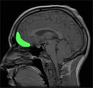
The orbitofrontal cortex (OFC) is a prefrontal cortex region in the frontal lobes of the brain which is involved in the cognitive process of decision-making. In non-human primates it consists of the association cortex areas Brodmann area 11, 12 and 13; in humans it consists of Brodmann area 10, 11 and 47.

The sensorimotor mu rhythm, also known as mu wave, comb or wicket rhythms or arciform rhythms, are synchronized patterns of electrical activity involving large numbers of neurons, probably of the pyramidal type, in the part of the brain that controls voluntary movement. These patterns as measured by electroencephalography (EEG), magnetoencephalography (MEG), or electrocorticography (ECoG), repeat at a frequency of 7.5–12.5 Hz, and are most prominent when the body is physically at rest. Unlike the alpha wave, which occurs at a similar frequency over the resting visual cortex at the back of the scalp, the mu rhythm is found over the motor cortex, in a band approximately from ear to ear. People suppress mu rhythms when they perform motor actions or, with practice, when they visualize performing motor actions. This suppression is called desynchronization of the wave because EEG wave forms are caused by large numbers of neurons firing in synchrony. The mu rhythm is even suppressed when one observes another person performing a motor action or an abstract motion with biological characteristics. Researchers such as V. S. Ramachandran and colleagues have suggested that this is a sign that the mirror neuron system is involved in mu rhythm suppression, although others disagree.

Manuel F. Casanova is the SmartState Endowed Chair in Childhood Neurotherapeutics and a professor of Biomedical Sciences at the University of South Carolina School of Medicine Greenville. He is a former Gottfried and Gisela Kolb Endowed Chair in Outpatient Psychiatry and a Professor of Anatomical Sciences and Neurobiology at the University of Louisville.

Teashirt homolog 3 is a protein that in humans is encoded by the TSHZ3 gene. In mice, it is a necessary part of the neural circuitry that controls breathing. The gene is also a homolog of the Drosophila melanogaster teashirt gene, which encodes a zinc finger transcription factor important for development of the trunk.

The evolution of the brain refers to the progressive development and complexity of neural structures over millions of years, resulting in the diverse range of brain sizes and functions observed across different species today, particularly in vertebrates.
Nancy Minshew is a Professor of Psychiatry and Neurology at the University of Pittsburgh. She directs the Center of Excellence in Autism Research and is an internationally known expert in the cognitive, neurological, and genetic bases of autism. Minshew was trained as a behavioral child neurologist, and she received an M.D. from the Washington University School of Medicine in St. Louis.
Gyrification is the process of forming the characteristic folds of the cerebral cortex. The peak of such a fold is called a gyrus, and its trough is called a sulcus. The neurons of the cerebral cortex reside in a thin layer of gray matter, only 2–4 mm thick, at the surface of the brain. Much of the interior volume is occupied by white matter, which consists of long axonal projections to and from the cortical neurons residing near the surface. Gyrification allows a larger cortical surface area, and hence greater cognitive functionality to fit inside a smaller cranium.
In psychology and neuroscience, executive dysfunction, or executive function deficit is a disruption to the efficacy of the executive functions, which is a group of cognitive processes that regulate, control, and manage other cognitive processes. Executive dysfunction can refer to both neurocognitive deficits and behavioural symptoms. It is implicated in numerous psychopathologies and mental disorders, as well as short-term and long-term changes in non-clinical executive control. Executive dysfunction is the mechanism underlying ADHD paralysis, and in a broader context, it can encompass other cognitive difficulties like planning, organizing, initiating tasks and regulating emotions. It is a core characteristic of ADHD and can elucidate numerous other recognized symptoms.
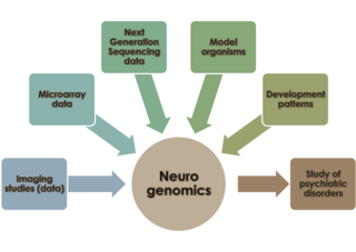
Neurogenomics is the study of how the genome of an organism influences the development and function of its nervous system. This field intends to unite functional genomics and neurobiology in order to understand the nervous system as a whole from a genomic perspective.
The causes of schizophrenia that underlie the development of schizophrenia, a psychiatric disorder, are complex and not clearly understood. A number of hypotheses including the dopamine hypothesis, and the glutamate hypothesis have been put forward in an attempt to explain the link between altered brain function and the symptoms and development of schizophrenia.
Autism spectrum disorder (ASD) refers to a variety of conditions typically identified by challenges with social skills, communication, speech, and repetitive sensory-motor behaviors. The 11th International Classification of Diseases (ICD-11), released in January 2021, characterizes ASD by the associated deficits in the ability to initiate and sustain two-way social communication and restricted or repetitive behavior unusual for the individual's age or situation. Although linked with early childhood, the symptoms can appear later as well. Symptoms can be detected before the age of two and experienced practitioners can give a reliable diagnosis by that age. However, official diagnosis may not occur until much older, even well into adulthood. There is a large degree of variation in how much support a person with ASD needs in day-to-day life. This can be classified by a further diagnosis of ASD level 1, level 2, or level 3. Of these, ASD level 3 describes people requiring very substantial support and who experience more severe symptoms. ASD-related deficits in nonverbal and verbal social skills can result in impediments in personal, family, social, educational, and occupational situations. This disorder tends to have a strong correlation with genetics along with other factors. More research is identifying ways in which epigenetics is linked to autism. Epigenetics generally refers to the ways in which chromatin structure is altered to affect gene expression. Mechanisms such as cytosine regulation and post-translational modifications of histones. Of the 215 genes contributing, to some extent in ASD, 42 have been found to be involved in epigenetic modification of gene expression. Some examples of ASD signs are specific or repeated behaviors, enhanced sensitivity to materials, being upset by changes in routine, appearing to show reduced interest in others, avoiding eye contact and limitations in social situations, as well as verbal communication. When social interaction becomes more important, some whose condition might have been overlooked suffer social and other exclusion and are more likely to have coexisting mental and physical conditions. Long-term problems include difficulties in daily living such as managing schedules, hypersensitivities, initiating and sustaining relationships, and maintaining jobs.
Cajal–Retzius cells are a heterogeneous population of morphologically and molecularly distinct reelin-producing cells. They are found in the marginal zone/layer I of the developing cerebral cortex and in the immature hippocampus of different species and at different times during embryogenesis and postnatal life.
The mechanisms of autism are the molecular and cellular processes believed to cause or contribute to the symptoms of autism. Multiple processes are hypothesized to explain different autism spectrum features. These hypotheses include defects in synapse structure and function, reduced synaptic plasticity, disrupted neural circuit function, gut–brain axis dyshomeostasis, neuroinflammation, and altered brain structure or connectivity. Autism symptoms stem from maturation-related changes in brain systems. The mechanisms of autism are divided into two main areas: pathophysiology of brain structures and processes, and neuropsychological linkages between brain structures and behaviors, with multiple pathophysiologies linked to various autism behaviors.

Karen Pierce is an American scientist known for her research on the early detection of autism spectrum disorder (ASD). She is a professor-in-residence in the Department of Neurosciences at University of California San Diego, and co-director of the UC San Diego Autism Center of Excellence (ACE). Pierce is an advocate of early detection and treatment engagement in autism spectrum disorder (ASD), supported by evidence of the brain’s high level of plasticity during early development.
References
- 1 2 Zimmer, Carl. "The Brain: The Troublesome Bloom of Autism". Discover Magazine. Retrieved 20 April 2013.
- ↑ Robbins, Gary. "UCSD finds genes possibly linked to autism". San Diego Union-Tribune. Retrieved 20 April 2013.
- 1 2 Jabr, Ferris. "The Ballooning Brain: Defective Genes May Explain Uncontrolled Brain Growth in Autism". Scientific American. Retrieved 20 April 2013.
- ↑ Hamilton, Teacha. "Excess of Neurons in Prefrontal Cortex Associated with Autism". Scicasts. Retrieved 20 April 2013.
- ↑ Park, Alice (9 November 2011). "Autistic Children Have More Brain Cells". Time. Retrieved 20 April 2013.
- ↑ Goodwin, Jennifer (8 November 2011). "Autistic children may have too many brain cells, study says". USA Today. Retrieved 20 April 2013.