
Functional magnetic resonance imaging or functional MRI (fMRI) measures brain activity by detecting changes associated with blood flow. This technique relies on the fact that cerebral blood flow and neuronal activation are coupled. When an area of the brain is in use, blood flow to that region also increases.

In haemodynamics, the body must respond to physical activities, external temperature, and other factors by homeostatically adjusting its blood flow to deliver nutrients such as oxygen and glucose to stressed tissues and allow them to function. Haemodynamic response (HR) allows the rapid delivery of blood to active neuronal tissues. The brain consumes large amounts of energy but does not have a reservoir of stored energy substrates. Since higher processes in the brain occur almost constantly, cerebral blood flow is essential for the maintenance of neurons, astrocytes, and other cells of the brain. This coupling between neuronal activity and blood flow is also referred to as neurovascular coupling.

A neural circuit is a population of neurons interconnected by synapses to carry out a specific function when activated. Multiple neural circuits interconnect with one another to form large scale brain networks.

The motion aftereffect (MAE) is a visual illusion experienced after viewing a moving visual stimulus for a time with stationary eyes, and then fixating a stationary stimulus. The stationary stimulus appears to move in the opposite direction to the original stimulus. The motion aftereffect is believed to be the result of motion adaptation.
Neural adaptation or sensory adaptation is a gradual decrease over time in the responsiveness of the sensory system to a constant stimulus. It is usually experienced as a change in the stimulus. For example, if a hand is rested on a table, the table's surface is immediately felt against the skin. Subsequently, however, the sensation of the table surface against the skin gradually diminishes until it is virtually unnoticeable. The sensory neurons that initially respond are no longer stimulated to respond; this is an example of neural adaptation.
Neural engineering is a discipline within biomedical engineering that uses engineering techniques to understand, repair, replace, or enhance neural systems. Neural engineers are uniquely qualified to solve design problems at the interface of living neural tissue and non-living constructs.

Neural oscillations, or brainwaves, are rhythmic or repetitive patterns of neural activity in the central nervous system. Neural tissue can generate oscillatory activity in many ways, driven either by mechanisms within individual neurons or by interactions between neurons. In individual neurons, oscillations can appear either as oscillations in membrane potential or as rhythmic patterns of action potentials, which then produce oscillatory activation of post-synaptic neurons. At the level of neural ensembles, synchronized activity of large numbers of neurons can give rise to macroscopic oscillations, which can be observed in an electroencephalogram. Oscillatory activity in groups of neurons generally arises from feedback connections between the neurons that result in the synchronization of their firing patterns. The interaction between neurons can give rise to oscillations at a different frequency than the firing frequency of individual neurons. A well-known example of macroscopic neural oscillations is alpha activity.

Neuronal noise or neural noise refers to the random intrinsic electrical fluctuations within neuronal networks. These fluctuations are not associated with encoding a response to internal or external stimuli and can be from one to two orders of magnitude. Most noise commonly occurs below a voltage-threshold that is needed for an action potential to occur, but sometimes it can be present in the form of an action potential; for example, stochastic oscillations in pacemaker neurons in suprachiasmatic nucleus are partially responsible for the organization of circadian rhythms.
Sensory neuroscience is a subfield of neuroscience which explores the anatomy and physiology of neurons that are part of sensory systems such as vision, hearing, and olfaction. Neurons in sensory regions of the brain respond to stimuli by firing one or more nerve impulses following stimulus presentation. How is information about the outside world encoded by the rate, timing, and pattern of action potentials? This so-called neural code is currently poorly understood and sensory neuroscience plays an important role in the attempt to decipher it. Looking at early sensory processing is advantageous since brain regions that are "higher up" contain neurons which encode more abstract representations. However, the hope is that there are unifying principles which govern how the brain encodes and processes information. Studying sensory systems is an important stepping stone in our understanding of brain function in general.

In vision, filling-in phenomena are those responsible for the completion of missing information across the physiological blind spot, and across natural and artificial scotomata. There is also evidence for similar mechanisms of completion in normal visual analysis. Classical demonstrations of perceptual filling-in involve filling in at the blind spot in monocular vision, and images stabilized on the retina either by means of special lenses, or under certain conditions of steady fixation. For example, naturally in monocular vision at the physiological blind spot, the percept is not a hole in the visual field, but the content is “filled-in” based on information from the surrounding visual field. When a textured stimulus is presented centered on but extending beyond the region of the blind spot, a continuous texture is perceived. This partially inferred percept is paradoxically considered more reliable than a percept based on external input..
Repetition priming refers to improvements in a behavioural response when stimuli are repeatedly presented. The improvements can be measured in terms of accuracy or reaction time, and can occur when the repeated stimuli are either identical or similar to previous stimuli. These improvements have been shown to be cumulative, so as the number of repetitions increases the responses get continually faster up to a maximum of around seven repetitions. These improvements are also found when the repeated items are changed slightly in terms of orientation, size and position. The size of the effect is also modulated by the length of time the item is presented for and the length time between the first and subsequent presentations of the repeated items.
Event-related functional magnetic resonance imaging (efMRI) is a technique used in magnetic resonance imaging of medical patients.

Biological neuron models, also known as a spiking neuron models, are mathematical descriptions of the properties of certain cells in the nervous system that generate sharp electrical potentials across their cell membrane, roughly one millisecond in duration, called action potentials or spikes. Since spikes are transmitted along the axon and synapses from the sending neuron to many other neurons, spiking neurons are considered to be a major information processing unit of the nervous system. Spiking neuron models can be divided into different categories: the most detailed mathematical models are biophysical neuron models that describe the membrane voltage as a function of the input current and the activation of ion channels. Mathematically simpler are integrate-and-fire models that describe the membrane voltage as a function of the input current and predict the spike times without a description of the biophysical processes that shape the time course of an action potential. Even more abstract models only predict output spikes as a function of the stimulation where the stimulation can occur through sensory input or pharmacologically. This article provides a short overview of different spiking neuron models and links, whenever possible to experimental phenomena. It includes deterministic and probabilistic models.
Neurophysics is the branch of biophysics dealing with the development and use of physical methods to gain information about the nervous system. Neurophysics is an interdisciplinary science using physics and combining it with other neurosciences to better understand neural processes. The methods used include the techniques of experimental biophysics and other physical measurements such as EEG mostly to study electrical, mechanical or fluidic properties, as well as theoretical and computational approaches. The term "neurophysics" is a portmanteau of "neuron" and "physics".
Synaptic noise refers to the constant bombardment of synaptic activity in neurons. This occurs in the background of a cell when potentials are produced without the nerve stimulation of an action potential, and are due to the inherently random nature of synapses. These random potentials have similar time courses as excitatory postsynaptic potentials (EPSPs) and inhibitory postsynaptic potentials (IPSPs), yet they lead to variable neuronal responses. The variability is due to differences in the discharge times of action potentials.

The neural correlates of consciousness (NCC) refer to the relationships between mental states and neural states and constitute the minimal set of neuronal events and mechanisms sufficient for a specific conscious percept. Neuroscientists use empirical approaches to discover neural correlates of subjective phenomena; that is, neural changes which necessarily and regularly correlate with a specific experience. The set should be minimal because, under the materialist assumption that the brain is sufficient to give rise to any given conscious experience, the question is which of its components are necessary to produce it.
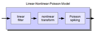
The linear-nonlinear-Poisson (LNP) cascade model is a simplified functional model of neural spike responses. It has been successfully used to describe the response characteristics of neurons in early sensory pathways, especially the visual system. The LNP model is generally implicit when using reverse correlation or the spike-triggered average to characterize neural responses with white-noise stimuli.
The H1 neuron is located in the visual cortex of true flies of the order Diptera and mediates motor responses to visual stimuli. H1 is sensitive to horizontal motion in the visual field and enables the fly to rapidly and accurately respond to optic flow with motor corrections to stabilize flight. It is particularly responsive to horizontal forward motion associated with movement of the fly's own body during flight. Damage to H1 impairs the fly's ability to counteract disturbances during flight, suggesting that it is a necessary component of the optomotor response. H1 is an ideal system for studying the neural basis of information processing due to its highly selective and predictable responses to stimuli. Since the initial anatomical and physiological characterizations of H1 in 1976, study of the neuron has greatly benefited the understanding of neural coding in a wide range of organisms, especially the relationship between the neural code and behavior.
The oddball paradigm is an experimental design used within psychology research. Presentations of sequences of repetitive stimuli are infrequently interrupted by a deviant stimulus. The reaction of the participant to this "oddball" stimulus is recorded.
The following outline is provided as an overview of and topical guide to brain mapping: