
The femoral artery is a large artery in the thigh and the main arterial supply to the thigh and leg. It enters the thigh from behind the inguinal ligament as the continuation of the external iliac artery.

The femoral triangle is an anatomical region of the upper third of the thigh. It is a subfascial space which appears as a triangular depression below the inguinal ligament when the thigh is flexed, abducted and laterally rotated.

The deep artery of the thigh, is a branch of the femoral artery that, as its name suggests, travels more deeply (posteriorly) than the rest of the femoral artery.

Legg–Calvé–Perthes disease (LCPD), is a childhood hip disorder initiated by a disruption of blood flow to the head of the femur. Due to the lack of blood flow, the bone dies and stops growing. Over time, healing occurs by new blood vessels infiltrating the dead bone and removing the necrotic bone which leads to a loss of bone mass and a weakening of the femoral head.

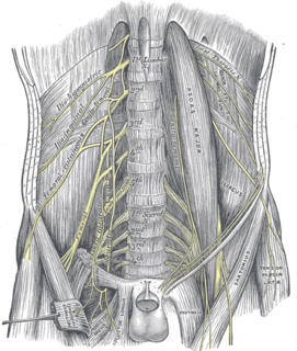
The genitofemoral nerve refers to a nerve that is found in the abdomen. Its branches, the genital branch and femoral branch supply sensation to the upper anterior thigh, as well as the skin of the anterior scrotum in males and mons veneris in females. The femoral branch is different from the femoral nerve, which also arises from the lumbar plexus.

The pectineus muscle is a flat, quadrangular muscle, situated at the anterior (front) part of the upper and medial (inner) aspect of the thigh. The pectineus muscle is the most anterior adductor of the hip. The muscle does adduct and medially rotate the thigh but its primary function is hip flexion.

The inguinal ligament, also known as Poupart's ligament or groin ligament, is a band running from the pubic tubercle to the anterior superior iliac spine. It forms the base of the inguinal canal through which an indirect inguinal hernia may develop.

In the human body, the femoral vein is a blood vessel that accompanies the femoral artery in the femoral sheath. Begins at the adductor hiatus and is a continuation of the popliteal vein. It ends at the inferior margin of the inguinal ligament, where it becomes the external iliac vein. The femoral vein bears valves which are mostly bicuspid and whose number is variable between individuals and often between left and right leg.

In vertebrate anatomy, hip refers to either an anatomical region or a joint.
In the human body, the adductor longus is a skeletal muscle located in the thigh. One of the adductor muscles of the hip, its main function is to adduct the thigh and it is innervated by the obturator nerve. It forms the medial wall of the femoral triangle.
The femoral nerve is a nerve in the thigh that supplies skin on the upper thigh and inner leg, and the muscles that extend the knee.

The lateral circumflex femoral artery is an artery in the upper thigh.
The medial circumflex femoral artery is an artery in the upper thigh that helps supply blood to the neck of the femur. Damage to the artery following a femoral neck fracture may lead to avascular necrosis (ischemic) of the femoral neck/head.

The adductor canal is an aponeurotic tunnel in the middle third of the thigh, extending from the apex of the femoral triangle to the opening in the adductor magnus, the adductor hiatus.
Femoral hernias occur just below the inguinal ligament, when abdominal contents pass through a naturally occurring weakness in the abdominal wall called the femoral canal. Femoral hernias are a relatively uncommon type, accounting for only 3% of all hernias. While femoral hernias can occur in both males and females, almost all develop in women due to the increased width of the female pelvis. Femoral hernias are more common in adults than in children. Those that do occur in children are more likely to be associated with a connective tissue disorder or with conditions that increase intra-abdominal pressure. Seventy percent of pediatric cases of femoral hernias occur in infants under the age of one.

The femoral ring is the base of the femoral canal. It is directed upward and is oval in form, its long diameter being directed transversely and measuring about 1.25 cm. Part of the intestine can sometimes pass through the femoral ring into the femoral canal causing a femoral hernia.

In human anatomy of the leg, the femoral sheath has three compartments. The lateral compartment contains the femoral artery, the intermediate compartment contains the femoral vein, and the medial and smallest compartment is called the femoral canal. The femoral canal contains efferent lymphatic vessels and a lymph node embedded in a small amount of areolar tissue. It is conical in shape and is about 2 cm long.
The cruciate anastomosis is a circulatory anastomosis in the upper thigh of the inferior gluteal artery, the lateral and medial circumflex femoral arteries, and the first perforating artery of the profunda femoris artery. Also, the anastomotic branch of the posterior branch of the obturator artery. The cruciate anastomosis is clinically relevant because if there is a blockage between the femoral artery and external iliac artery, blood can reach the popliteal artery by means of the anastomosis. The route of blood is through the internal iliac, to the inferior gluteal artery, to a perforating branch of the deep femoral artery, to the lateral circumflex femoral artery, then to its descending branch into the superior lateral genicular artery and thus into the popliteal artery.
The femoral sheath is formed by a prolongation downward, behind the inguinal ligament, of the abdominal fascia, the transverse fascia being continued down in front of the femoral vessels and the iliac fascia behind them. The femoral sheath is contained within the femoral triangle.

The femoral head is the highest part of the thigh bone (femur). It is supported by the femoral neck.