Fetal head

Last updated

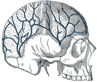
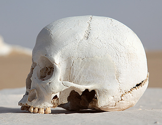
The fetal head, from an obstetrical viewpoint, and in particular its size, is important because an essential feature of labor is the adaptation between the fetal head and the maternal bony pelvis. Only a comparatively small part of the head at term is represented by the face. The rest of the head is composed of the firm skull, which is made up of two frontal, two parietal, and two temporal bones, along with the upper portion of the occipital bone and the wings of the sphenoid.

Pelvis lower part of the trunk of the human body between the abdomen and the thighs (sometimes also called pelvic region of the trunk

The pelvis is either the lower part of the trunk of the human body between the abdomen and the thighs or the skeleton embedded in it.

Temporal bone bones situated at the sides and base of the skull, and lateral to the temporal lobes of the cerebrum

The temporal bones are situated at the sides and base of the skull, and lateral to the temporal lobes of the cerebral cortex.

Sphenoid bone unpaired bone situated at the front middle of the skull in front of the temporal bone and basilar part of the occipital bone

The sphenoid bone is an unpaired bone of the neurocranium. It is situated in the middle of the skull towards the front, in front of the basilar part of the occipital bone. The sphenoid bone is one of the seven bones that articulate to form the orbit. Its shape somewhat resembles that of a butterfly or bat with its wings extended.

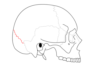
These bones are separated by membranous spaces, or sutures. The most important sutures are the frontal, between the two frontal bones; the sagittal, between the two parietal bones; the two coronal, between the frontal and parietal bones; and the two lambdoid, between the posterior margins of the parietal bones and upper margin of the occipital bone. Where several sutures meet, an irregular space forms, which is enclosed by a membrane and designated as a fontanel. The greater, or anterior fontanel, is a lozenge-shaped space that is situated at the junction of the sagittal and the coronal sutures. The lesser, or posterior fontanel, is represented by a small triangular area at the intersection of the sagittal and lambdoid sutures. The localization of these fontanels gives important information concerning the presentation and position of the fetus. The temporal, or casserian fontanels, have no diagnostic

Parietal bone bone in the human skull which, when joined together, forms the sides and roof of the cranium

The parietal bones are two bones in the skull which, when joined together at a fibrous joint, form the sides and roof of the cranium. In humans, each bone is roughly quadrilateral in form, and has two surfaces, four borders, and four angles. It is named from the Latin paries (-ietis), wall.

Occipital bone saucer-shaped membrane bone situated at the back and lower part of the cranium

The occipital bone is a cranial dermal bone and the main bone of the occiput. It is trapezoidal in shape and curved on itself like a shallow dish. The occipital bone overlies the occipital lobes of the cerebrum. At the base of skull in the occipital bone, there is a large oval opening called the foramen magnum, which allows the passage of the spinal cord.

It is customary to measure certain critical diameters and circumferences of the newborn head. The diameters most frequently used, and the average lengths thereof, are:

  1. The occipitofrontal (11.5 cm), which follows a line extending from a point just above the root of the nose to the most prominent portion of the occipital bone.
  2. The biparietal (9.5 cm), the greatest transverse diameter of the head, which extends from one parietal boss to the other.
  3. The bitemporal (8.0 cm), the greatest distance between the two temporal sutures.
  4. The occipitomental (12.5 cm), from the chin to the most prominent portion of the occiput.
  5. The suboccipitobregmatic (9.5 cm), which follows a line drawn from the middle of the large fontanel to the undersurface of the occipital bone just where it joins the neck.

The greatest circumference of the head, which corresponds to the plane of the occipitofrontal diameter, averages 34.5 cm, a size too large to fit through the pelvis without flexion. The smallest circumference, corresponding to the plane of the suboccipitobregmatic diameter, is 32 cm. The bones of the cranium are normally connected only by a thin layer of fibrous tissue that allows considerable shifting or sliding of each bone to accommodate the size and shape of the maternal pelvis. This intrapartum process is termed molding. The head position and degree of skull ossification result in a spectrum of cranial plasticity from minimal to great and in some cases, undoubtedly contribute to fetopelvic disproportion, a leading indication for cesarean delivery.

Related Research Articles

Skull bony structure that forms the skeleton of head in most vertebrates

The skull is a bony structure that forms the head in vertebrates. It supports the structures of the face and provides a protective cavity for the brain. The skull is composed of two parts: the cranium and the mandible. In humans, these two parts are the neurocranium and the viscerocranium or facial skeleton that includes the mandible as its largest bone. The skull forms the anterior most portion of the skeleton and is a product of cephalisation—housing the brain, and several sensory structures such as the eyes, ears, nose, and mouth. In humans these sensory structures are part of the facial skeleton.

Fontanelle membrane connecting parts of a babys skull

A fontanelle is an anatomical feature of the infant human skull comprising any of the soft membranous gaps (sutures) between the cranial bones that make up the calvaria of a fetus or an infant. Fontanelles allow for rapid stretching and deformation of the neurocranium as the brain expands faster than the surrounding bone can grow. Premature complete ossification of the sutures is called craniosynostosis.

Craniosynostosis synostosis that results in premature fusion located in skull

Craniosynostosis is a condition in which one or more of the fibrous sutures in an infant skull prematurely fuses by turning into bone (ossification), thereby changing the growth pattern of the skull. Because the skull cannot expand perpendicular to the fused suture, it compensates by growing more in the direction parallel to the closed sutures. Sometimes the resulting growth pattern provides the necessary space for the growing brain, but results in an abnormal head shape and abnormal facial features. In cases in which the compensation does not effectively provide enough space for the growing brain, craniosynostosis results in increased intracranial pressure leading possibly to visual impairment, sleeping impairment, eating difficulties, or an impairment of mental development combined with a significant reduction in IQ.

Diploic veins

The diploic veins are large, thin-walled valveless veins that channel in the diploë between the inner and outer layers of the cortical bone in the skull. They are lined by a single layer of endothelium supported by elastic tissue. They develop fully by the age of two years. The diploic veins drain this area into the dural venous sinuses. The four major trunks of the diploic veins found on each side of the head are frontal, anterior temporal, posterior temporal, and occipital diploic veins.

Sagittal suture

The sagittal suture is a dense, fibrous connective tissue joint between the two parietal bones of the skull. The term is derived from the Latin word sagitta, meaning arrow. The derivation of this term may be demonstrated by observing how the sagittal suture is notched posteriorly, like an arrow, by the lambdoid suture. The sagittal suture is also known as the interparietal suture, the sutura interparietalis.

Lambdoid suture

The lambdoid suture is a dense, fibrous connective tissue joint on the posterior aspect of the skull that connects the parietal bones with the occipital bone. It is continuous with the occipitomastoid suture.

Coronal suture

The coronal suture is a dense, fibrous connective tissue joint that separates the two parietal bones from the frontal bone of the skull.

Asterion (anatomy)

The asterion is the point on the skull corresponding to the posterior end of the parietomastoid suture.

<i>Galesaurus</i> genus of mammals (fossil)

Galesaurus was a prehistoric carnivorous therapsid that lived between the Induan and the Olenekian age in what is now South Africa. It was incorrectly classified as a dinosaur by Sir Richard Owen in 1859.

Superior sagittal sinus

The superior sagittal sinus, within the human head, is an unpaired area along the attached margin of the falx cerebri. It allows blood to drain from the lateral aspects of anterior cerebral hemispheres to the confluence of sinuses. Cerebrospinal fluid drains through arachnoid granulations into the superior sagittal sinus and is returned to venous circulation.

Transverse sinuses

The transverse sinuses, within the human head, are two areas beneath the brain which allow blood to drain from the back of the head. They run laterally in a groove along the interior surface of the occipital bone. They drain from the confluence of sinuses to the sigmoid sinuses, which ultimately connect to the internal jugular vein. See diagram : labeled under the brain as "SIN. TRANS.".

Pterion

The pterion is the region where the frontal, parietal, temporal, and sphenoid bones join together. It is located on the side of the skull, just behind the temple.

Anterior fontanelle

The anterior fontanelle is the largest fontanelle, and is placed at the junction of the sagittal suture, coronal suture, and frontal suture; it is lozenge-shaped, and measures about 4 cm in its antero-posterior and 2.5 cm in its transverse diameter. The fontanelle allows the skull to deform during birth to ease its passage through the birth canal and for expansion of the brain after birth.

Fibrous joint

Fibrous joints are connected by dense connective tissue, consisting mainly of collagen. These are fixed joints where bones are united by a layer of white fibrous tissue of varying thickness. In the skull the joints between the bones are called sutures. Such immovable joints are also referred to as synarthroses.

Calvaria (skull) upper part of the neurocranium that covers the cranial cavity containing the brain

The calvaria or skullcap is the top part of the skull. It is the upper part of the neurocranium and covers the cranial cavity containing the brain. It forms the main component of the skull roof.

In arthropod and vertebrate anatomy, the vertex is the upper surface of the head.

McGillivray syndrome Familial scaphocephaly syndrome, McGillivray type is a rare newly described craniosynostosis (see this term) syndrome characterized by scaphocephaly, macrocephaly, severe maxillary retrusion, and mild intellectual disability

McGillivray syndrome is a rare syndrome characterized mainly by heart defects, skull and facial abnormalities and ambiguous genitalia. The symptoms of this syndrome are ventricular septal defect, patent ductus arteriosus, small jaw, undescended testes, and webbed fingers. Beside to these symptoms there are more symptoms which is related with bone structure and misshape.

References

Cunningham, F. Gary; Leveno, Kenneth J.; Gilstrap, Larry C. III; Hauth, John C. (March 31, 2005). Williams Obstetrics (22nd ed.). McGraw-Hill Professional. ISBN   978-0071413152.

International Standard Book Number Unique numeric book identifier

The International Standard Book Number (ISBN) is a numeric commercial book identifier which is intended to be unique. Publishers purchase ISBNs from an affiliate of the International ISBN Agency.