Related Research Articles

A laser is a device that emits light through a process of optical amplification based on the stimulated emission of electromagnetic radiation. The word laser is an anacronym that originated as an acronym for light amplification by stimulated emission of radiation. The first laser was built in 1960 by Theodore Maiman at Hughes Research Laboratories, based on theoretical work by Charles H. Townes and Arthur Leonard Schawlow.
An attosecond is a unit of time in the International System of Units (SI) equal to 10−18 or 1⁄1 000 000 000 000 000 000 of a second. An attosecond is to a second as a second is to about 31.71 billion years. The attosecond is a newly discovered "slice of time" that is tiny but has various potential applications: it can observe oscillating molecules, the chemical bonds formed by atoms in chemical reactions, and other extremely tiny and extremely fast things.

Femtochemistry is the area of physical chemistry that studies chemical reactions on extremely short timescales in order to study the very act of atoms within molecules (reactants) rearranging themselves to form new molecules (products). In a 1988 issue of the journal Science, Ahmed Hassan Zewail published an article using this term for the first time, stating "Real-time femtochemistry, that is, chemistry on the femtosecond timescale...". Later in 1999, Zewail received the Nobel Prize in Chemistry for his pioneering work in this field showing that it is possible to see how atoms in a molecule move during a chemical reaction with flashes of laser light.
In physics and physical chemistry, time-resolved spectroscopy is the study of dynamic processes in materials or chemical compounds by means of spectroscopic techniques. Most often, processes are studied after the illumination of a material occurs, but in principle, the technique can be applied to any process that leads to a change in properties of a material. With the help of pulsed lasers, it is possible to study processes that occur on time scales as short as 10−16 seconds. All time-resolved spectra are suitable to be analyzed using the two-dimensional correlation method for a correlation map between the peaks.
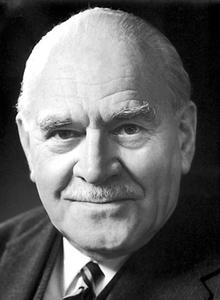
Ronald George Wreyford Norrish FRS was a British chemist who was awarded the Nobel Prize in Chemistry in 1967.

George Porter, Baron Porter of Luddenham, was a British chemist. He was awarded the Nobel Prize in Chemistry in 1967.
Coherent anti-Stokes Raman spectroscopy, also called Coherent anti-Stokes Raman scattering spectroscopy (CARS), is a form of spectroscopy used primarily in chemistry, physics and related fields. It is sensitive to the same vibrational signatures of molecules as seen in Raman spectroscopy, typically the nuclear vibrations of chemical bonds. Unlike Raman spectroscopy, CARS employs multiple photons to address the molecular vibrations, and produces a coherent signal. As a result, CARS is orders of magnitude stronger than spontaneous Raman emission. CARS is a third-order nonlinear optical process involving three laser beams: a pump beam of frequency ωp, a Stokes beam of frequency ωS and a probe beam at frequency ωpr. These beams interact with the sample and generate a coherent optical signal at the anti-Stokes frequency (ωpr+ωp-ωS). The latter is resonantly enhanced when the frequency difference between the pump and the Stokes beams (ωp-ωS) coincides with the frequency of a Raman resonance, which is the basis of the technique's intrinsic vibrational contrast mechanism.
Radiation chemistry is a subdivision of nuclear chemistry which studies the chemical effects of ionizing radiation on matter. This is quite different from radiochemistry, as no radioactivity needs to be present in the material which is being chemically changed by the radiation. An example is the conversion of water into hydrogen gas and hydrogen peroxide.
Photodissociation, photolysis, photodecomposition, or photofragmentation is a chemical reaction in which molecules of a chemical compound are broken down by photons. It is defined as the interaction of one or more photons with one target molecule.
Ultrafast laser spectroscopy is a spectroscopic technique that uses ultrashort pulse lasers for the study of dynamics on extremely short time scales. Different methods are used to examine the dynamics of charge carriers, atoms, and molecules. Many different procedures have been developed spanning different time scales and photon energy ranges; some common methods are listed below.
The European X-Ray Free-Electron Laser Facility is an X-ray research laser facility commissioned during 2017. The first laser pulses were produced in May 2017 and the facility started user operation in September 2017. The international project with twelve participating countries; nine shareholders at the time of commissioning, later joined by three other partners, is located in the German federal states of Hamburg and Schleswig-Holstein. A free-electron laser generates high-intensity electromagnetic radiation by accelerating electrons to relativistic speeds and directing them through special magnetic structures. The European XFEL is constructed such that the electrons produce X-ray light in synchronisation, resulting in high-intensity X-ray pulses with the properties of laser light and at intensities much brighter than those produced by conventional synchrotron light sources.
In analytical chemistry, crossed molecular beam experiments involve two beams of atoms or molecules which are collided together to study the dynamics of the chemical reaction, and can detect individual reactive collisions.
Photoelectrochemical processes are processes in photoelectrochemistry; they usually involve transforming light into other forms of energy. These processes apply to photochemistry, optically pumped lasers, sensitized solar cells, luminescence, and photochromism.

Nuclear magnetic resonance (NMR) is a physical phenomenon in which nuclei in a strong constant magnetic field are perturbed by a weak oscillating magnetic field and respond by producing an electromagnetic signal with a frequency characteristic of the magnetic field at the nucleus. This process occurs near resonance, when the oscillation frequency matches the intrinsic frequency of the nuclei, which depends on the strength of the static magnetic field, the chemical environment, and the magnetic properties of the isotope involved; in practical applications with static magnetic fields up to ca. 20 tesla, the frequency is similar to VHF and UHF television broadcasts (60–1000 MHz). NMR results from specific magnetic properties of certain atomic nuclei. Nuclear magnetic resonance spectroscopy is widely used to determine the structure of organic molecules in solution and study molecular physics and crystals as well as non-crystalline materials. NMR is also routinely used in advanced medical imaging techniques, such as in magnetic resonance imaging (MRI). The original application of NMR to condensed matter physics is nowadays mostly devoted to strongly correlated electron systems. It reveals large manybody couplings by fast broadband detection and it should not to be confused with solid state NMR, which aims at removing the effect of the same couplings by Magic Angle Spinning techniques.
Time resolved crystallography utilizes X-ray crystallography imaging to visualize reactions in four dimensions. This enables the studies of dynamical changes that occur in for example enzymes during their catalysis. The time dimension is incorporated by triggering the reaction of interest in the crystal prior to X-ray exposure, and then collecting the diffraction patterns at different time delays. In order to study these dynamical properties of macromolecules three criteria must be met;
The photoacoustic effect or optoacoustic effect is the formation of sound waves following light absorption in a material sample. In order to obtain this effect the light intensity must vary, either periodically or as a single flash. The photoacoustic effect is quantified by measuring the formed sound with appropriate detectors, such as microphones or piezoelectric sensors. The time variation of the electric output from these detectors is the photoacoustic signal. These measurements are useful to determine certain properties of the studied sample. For example, in photoacoustic spectroscopy, the photoacoustic signal is used to obtain the actual absorption of light in either opaque or transparent objects. It is useful for substances in extremely low concentrations, because very strong pulses of light from a laser can be used to increase sensitivity and very narrow wavelengths can be used for specificity. Furthermore, photoacoustic measurements serve as a valuable research tool in the study of the heat evolved in photochemical reactions, particularly in the study of photosynthesis.

Albert Stolow is a Canadian physicist. He is the Canada Research Chair in Molecular Photonics, full professor of chemistry & biomolecular sciences and of physics, and a member of the Ottawa Institute for Systems Biology at the University of Ottawa. He is the founder and an ongoing member of the Molecular Photonics Group at the National Research Council of Canada. He is adjunct professor of Chemistry and of Physics at Queen's University in Kingston, and a Graduate Faculty Scholar in the department of physics, University of Central Florida and a Fellow of the Max-Planck-uOttawa Centre for Extreme and Quantum Photonics. In 2008, he was elected a Fellow in the American Physical Society, nominated by its Division of Chemical Physics in 2008, for contributions to ultrafast laser science as applied to molecular physics, including time-resolved studies of non-adiabatic dynamics in excited molecules, non-perturbative quantum control of molecular dynamics, and dynamics of polyatomic molecules in strong laser fields. In 2008, Stolow won the Keith Laidler Award of the Canadian Society for Chemistry, for a distinguished contribution to the field of physical chemistry, recognizing early career achievement. In 2009, he was elected a Fellow of the Optical Society of America for the application of ultrafast optical techniques to molecular dynamics and control, in particular, studies of molecules in strong laser fields and the development of new methods of optical quantum control. In 2013, he was awarded the Queen Elizabeth II Diamond Jubilee Medal (Canada). In 2017, Stolow was awarded the Earle K. Plyler Prize for Molecular Spectroscopy and Dynamics of the American Physical Society for the development of methods for probing and controlling ultrafast dynamics in polyatomic molecules, including time-resolved photoelectron spectroscopy and imaging, strong field molecular ionization, and dynamic Stark quantum control. In 2018, Stolow was awarded the John C. Polanyi Award of the Canadian Society for Chemistry “for excellence by a scientist carrying out research in Canada in physical, theoretical or computational chemistry or chemical physics”. In 2020, he became Chair of the Division of Chemical Physics of the American Physical Society. His group's research interests include ultrafast molecular dynamics and quantum control, time-resolved photoelectron spectroscopy and imaging, strong field & attosecond physics of polyatomic molecules, and coherent non-linear optical microscopy of live cells/tissues, materials and geological samples. In 2020, Stolow launched a major new high power ultrafast laser facility at the University of Ottawa producing high energy, phase-controlled few-cycle pulses of 2 micron wavelength at 10 kHz repetition rate. These are used for High Harmonic Generation to produce bright ultrafast Soft X-ray pulses for a new Ultrafast Xray Science Laboratory.
Amy S. Mullin is an American chemist and professor at the University of Maryland. She is a Fellow of the American Physical Society, the American Association for the Advancement of Science and the Optical Society of America. Her research focuses on molecular dynamics.
Ultrafast scanning electron microscopy (UFSEM) combines two microscopic modalities, Pump-probe microscopy and Scanning electron microscope, to gather temporal and spatial resolution phenomena. The technique uses ultrashort laser pulses for pump excitation of the material and the sample response will be detected by an Everhart-Thornley detector. Acquiring data depends mainly on formation of images by raster scan mode after pumping with short laser pulse at different delay times. The characterization of the output image will be done through the temporal resolution aspect. Thus, the idea is to exploit the shorter DeBroglie wavelength in respect to the photons which has great impact to increase the resolution about 1 nm. That technique is an up-to-date approach to study the dynamic of charge on material surfaces.
References
- ↑ "Laser flash photolysis". www.rsc.org. Royal Society of Chemistry. Retrieved 7 January 2024.
- ↑ "The Nobel Prize in Chemistry 1967". Nobelprize.org. 1967. Retrieved 20 March 2018.
| International | |
|---|---|
| National | |