Fluorescent D-amino acids

Last updated

Fluorescent D-amino acids (FDAAs) are D-amino acid derivatives whose side-chain terminal is covalently coupled with a fluorophore molecule. [1] FDAAs incorporate into the bacterial peptidoglycan (PG) in live bacteria, resulting in strong peripheral and septal PG labeling without affecting cell growth. They are featured with their in-situ incorporation mechanisms which enable time-course tracking of new PG formation. [2] To date, FDAAs have been employed for studying the cell wall synthesis in various bacterial species (both Gram-positives and Gram-negatives) through different techniques, such as microscopy, mass spectrometry, flow cytometry.

Contents

Structures and general properties

Collection of reported fluorescent D-amino acids and their structures. FDAA structures.jpg
Collection of reported fluorescent D-amino acids and their structures.

FDAA consists of a D-amino acid and a fluorophore (coupled through the amino acid side chain.) The D-amino acid backbone is required for its incorporation into the bacterial peptidoglycan through the activity of DD-transpeptidases. [3] Once being incorporated, one can use fluorescence-detection techniques to visualize the location of new PG formation as well as the growth rate. [4]

D-alanine is the most well-studied D-amino acids for FDAA development because it is a naturally existing residue in bacterial peptidoglycan structures. On the other hand, various fluorophores have been employed for FDAA applications and each has its features. [5] For example, coumarin-based FDAA (HADA) is small enough to penetrate the bacterial outer-membranes and thus is widely used for Gram-negative bacterial studies; while TAMRA-based FDAA (TADA) features its high brightness and photo/thermo-stability, which is suitable for super-resolution microscopy (strong excitation light is used). [5]

Proposed FDAA incorporation mechanisms

Proposed mechanism of FDAA incorporation into bacterial peptidoglycan. Mechanism of FDAA labeling.png
Proposed mechanism of FDAA incorporation into bacterial peptidoglycan.

Peptidoglycan (PG) is a mesh-like structure containing polysaccharides cross-linked by peptide chains. [6] Penicillin-binding proteins (DD-transpeptidases), in short PBPs, recognize the PG peptides and catalyze the cross-linking reactions. [7] These enzymes are reported to have high specificity toward the chirality center of the amino acid backbone (the D-chiral center) but relatively low specificity toward the side-chain structure. Therefore, when FDAAs are present, they are taken by PBPs for the cross-linking reactions, resulting in their incorporation into the PG peptide chains. At proper concentration, e.g. 1-2 mM, FDAAs labeling does not affect PG synthesis and cell growth because only 1-2% of PG peptide chains are labeled with FDAA. [2]

Sequential labeling of FDAAs revealed the growth pattern of peptidoglycan in S. venezuelae. Sequential labeling of S venezuelae.tif
Sequential labeling of FDAAs revealed the growth pattern of peptidoglycan in S. venezuelae.

Applications

Published studies utilizing FDAAs as tools include:

Related Research Articles

Peptidoglycan or murein is a unique large macromolecule, a polysaccharide, consisting of sugars and amino acids that forms a mesh-like peptidoglycan layer outside the plasma membrane, the rigid cell wall characteristic of most bacteria. The sugar component consists of alternating residues of β-(1,4) linked N-acetylglucosamine (NAG) and N-acetylmuramic acid (NAM). Attached to the N-acetylmuramic acid is an oligopeptide chain made of three to five amino acids. The peptide chain can be cross-linked to the peptide chain of another strand forming the 3D mesh-like layer. Peptidoglycan serves a structural role in the bacterial cell wall, giving structural strength, as well as counteracting the osmotic pressure of the cytoplasm. This repetitive linking results in a dense peptidoglycan layer which is critical for maintaining cell form and withstanding high osmotic pressures, and it is regularly replaced by peptidoglycan production. Peptidoglycan hydrolysis and synthesis are two processes that must occur in order for cells to grow and multiply, a technique carried out in three stages: clipping of current material, insertion of new material, and re-crosslinking of existing material to new material.

Autolysins are endogenous lytic enzymes that break down the peptidoglycan components of biological cells which enables the separation of daughter cells following cell division. They are involved in cell growth, cell wall metabolism, cell division and separation, as well as peptidoglycan turnover and have similar functions to lysozymes.

<span class="mw-page-title-main">Beta-lactam antibiotics</span> Class of broad-spectrum antibiotics

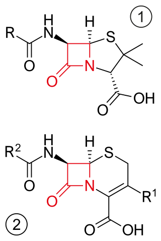
β-lactam antibiotics are antibiotics that contain a beta-lactam ring in their chemical structure. This includes penicillin derivatives (penams), cephalosporins and cephamycins (cephems), monobactams, carbapenems and carbacephems. Most β-lactam antibiotics work by inhibiting cell wall biosynthesis in the bacterial organism and are the most widely used group of antibiotics. Until 2003, when measured by sales, more than half of all commercially available antibiotics in use were β-lactam compounds. The first β-lactam antibiotic discovered, penicillin, was isolated from a strain of Penicillium rubens.

Braun's lipoprotein, found in some gram-negative cell walls, is one of the most abundant membrane proteins; its molecular weight is about 7.2 kDa. It is bound at its C-terminal end by a covalent bond to the peptidoglycan layer and is embedded in the outer membrane by its hydrophobic head. BLP tightly links the two layers and provides structural integrity to the outer membrane.

<span class="mw-page-title-main">DD-transpeptidase</span>

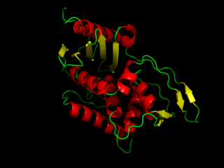
DD-transpeptidase is a bacterial enzyme that catalyzes the transfer of the R-L-αα-D-alanyl moiety of R-L-αα-D-alanyl-D-alanine carbonyl donors to the γ-OH of their active-site serine and from this to a final acceptor. It is involved in bacterial cell wall biosynthesis, namely, the transpeptidation that crosslinks the peptide side chains of peptidoglycan strands.

<i>N</i>-Acetylmuramic acid Chemical compound

N-Acetylmuramic acid is an organic compound with the chemical formula C
11
H
19
NO
8
. It is a monomer of peptidoglycan in most bacterial cell walls, which is built from alternating units of N-acetylglucosamine (GlcNAc) and N-acetylmuramic acid, cross-linked by oligopeptides at the lactic acid residue of MurNAc.

<span class="mw-page-title-main">Glycopeptide antibiotic</span> Class of antibiotic drugs

Glycopeptide antibiotics are a class of drugs of microbial origin that are composed of glycosylated cyclic or polycyclic nonribosomal peptides. Significant glycopeptide antibiotics include the anti-infective antibiotics vancomycin, teicoplanin, telavancin, ramoplanin and decaplanin, corbomycin, complestatin and the antitumor antibiotic bleomycin. Vancomycin is used if infection with methicillin-resistant Staphylococcus aureus (MRSA) is suspected.

<span class="mw-page-title-main">Pseudopeptidoglycan</span>

Pseudopeptidoglycan is a major cell wall component of some Archaea that differs from bacterial peptidoglycan in chemical structure, but resembles bacterial peptidoglycan in function and physical structure. Pseudopeptidoglycan, in general, is only present in a few methanogenic archaea. The basic components are N-acetylglucosamine and N-acetyltalosaminuronic acid, which are linked by β-1,3-glycosidic bonds.

<span class="mw-page-title-main">Penicillin-binding proteins</span> Class of proteins

Penicillin-binding proteins (PBPs) are a group of proteins that are characterized by their affinity for and binding of penicillin. They are a normal constituent of many bacteria; the name just reflects the way by which the protein was discovered. All β-lactam antibiotics bind to PBPs, which are essential for bacterial cell wall synthesis. PBPs are members of a subgroup of enzymes called transpeptidases. Specifically, PBPs are DD-transpeptidases.

<span class="mw-page-title-main">Lysin</span>

Lysins, also known as endolysins or murein hydrolases, are hydrolytic enzymes produced by bacteriophages in order to cleave the host's cell wall during the final stage of the lytic cycle. Lysins are highly evolved enzymes that are able to target one of the five bonds in peptidoglycan (murein), the main component of bacterial cell walls, which allows the release of progeny virions from the lysed cell. Cell-wall-containing Archaea are also lysed by specialized pseudomurein-cleaving lysins, while most archaeal viruses employ alternative mechanisms. Similarly, not all bacteriophages synthesize lysins: some small single-stranded DNA and RNA phages produce membrane proteins that activate the host's autolytic mechanisms such as autolysins.

In enzymology, glutamate racemase is an enzyme that catalyzes the chemical reaction

Peptidoglycan glycosyltransferase is an enzyme used in the biosynthesis of peptidoglycan. It transfers a disaccharide-peptide from a donor substrate to synthesize a glycan chain.

Zinc D-Ala-D-Ala carboxypeptidase (EC 3.4.17.14, Zn2+ G peptidase, D-alanyl-D-alanine hydrolase, D-alanyl-D-alanine-cleaving carboxypeptidase, DD-carboxypeptidase, G enzyme, DD-carboxypeptidase-transpeptidase) is an enzyme. This enzyme catalyses the following chemical reaction

Sortases are membrane anchored enzyme that sort these surface proteins onto the bacterial cell surface and anchor them to the peptidoglycan. There are different types of sortases and each catalyse the anchoring of different proteins to cell walls.

<span class="mw-page-title-main">Lipid II</span> Chemical compound

Lipid II is a precursor molecule in the synthesis of the cell wall of bacteria. It is a peptidoglycan, which is amphipathic and named for its bactoprenol hydrocarbon chain, which acts as a lipid anchor, embedding itself in the bacterial cell membrane. Lipid II must translocate across the cell membrane to deliver and incorporate its disaccharide-pentapeptide "building block" into the peptidoglycan mesh. Lipid II is the target of several antibiotics.

<span class="mw-page-title-main">Divisome</span> A protein complex in bacteria responsible for cell division

The divisome is a protein complex in bacteria that is responsible for cell division, constriction of inner and outer membranes during division, and peptidoglycan (PG) synthesis at the division site. The divisome is a membrane protein complex with proteins on both sides of the cytoplasmic membrane. In gram-negative cells it is located in the inner membrane. The divisome is nearly ubiquitous in bacteria although its composition may vary between species.

Renee Elizabeth Sockett is a professor and microbiologist in the School of Life Sciences at the University of Nottingham. She is a world-leading expert on Bdellovibrio bacteriovorus, a species of predatory bacteria.

D-Amino acids are amino acids where the stereogenic carbon alpha to the amino group has the D-configuration. For most naturally-occurring amino acids, this carbon has the L-configuration. D-Amino acids are occasionally found in nature as residues in proteins. They are formed from ribosomally-derived D-amino acid residues.

Corbomycin is a member of the glycopeptide family of antibiotics that are produced by soil bacteria.

<span class="mw-page-title-main">Joshua Shaevitz</span> American biophysicist

Joshua Shaevitz is an American biophysicist and Professor of Physics at the Lewis-Sigler Institute at Princeton University in Princeton, NJ. He is known for his work in single-molecule biophysics, bacterial growth and motility, and animal behavior.

References

  1. 1 2 3 4 Hsu, Yen-Pang; Booher, Garrett; Egan, Alexander; Vollmer, Waldemar; VanNieuwenhze, Michael S. (2019-09-17). "d -Amino Acid Derivatives as in Situ Probes for Visualizing Bacterial Peptidoglycan Biosynthesis". Accounts of Chemical Research. 52 (9): 2713–2722. doi:10.1021/acs.accounts.9b00311. ISSN   0001-4842. PMID   31419110. S2CID   206385813.
  2. 1 2 3 Kuru, Erkin; Hughes, H. Velocity; Brown, Pamela J.; Hall, Edward; Tekkam, Srinivas; Cava, Felipe; de Pedro, Miguel A.; Brun, Yves V.; VanNieuwenhze, Michael S. (2012-12-07). "In Situ Probing of Newly Synthesized Peptidoglycan in Live Bacteria with Fluorescent D -Amino Acids". Angewandte Chemie International Edition. 51 (50): 12519–12523. doi:10.1002/anie.201206749. PMC   3589519 . PMID   23055266.
  3. Kuru, Erkin; Radkov, Atanas; Meng, Xin; Egan, Alexander; Alvarez, Laura; Dowson, Amanda; Booher, Garrett; Breukink, Eefjan; Roper, David I.; Cava, Felipe; Vollmer, Waldemar (2019-12-20). "Mechanisms of Incorporation for D -Amino Acid Probes That Target Peptidoglycan Biosynthesis". ACS Chemical Biology. 14 (12): 2745–2756. doi:10.1021/acschembio.9b00664. ISSN   1554-8929. PMC   6929685 . PMID   31743648.
  4. 1 2 Radkov, Atanas D.; Hsu, Yen-Pang; Booher, Garrett; VanNieuwenhze, Michael S. (2018-06-20). "Imaging Bacterial Cell Wall Biosynthesis". Annual Review of Biochemistry. 87 (1): 991–1014. doi:10.1146/annurev-biochem-062917-012921. ISSN   0066-4154. PMC   6287495 . PMID   29596002.
  5. 1 2 Hsu, Yen-Pang; Rittichier, Jonathan; Kuru, Erkin; Yablonowski, Jacob; Pasciak, Erick; Tekkam, Srinivas; Hall, Edward; Murphy, Brennan; Lee, Timothy K.; Garner, Ethan C.; Huang, Kerwyn Casey (2017). "Full color palette of fluorescent d -amino acids for in situ labeling of bacterial cell walls". Chemical Science. 8 (9): 6313–6321. doi:10.1039/C7SC01800B. ISSN   2041-6520. PMC   5628581 . PMID   28989665.
  6. Vollmer, Waldemar; Blanot, Didier; De Pedro, Miguel A. (March 2008). "Peptidoglycan structure and architecture". FEMS Microbiology Reviews. 32 (2): 149–167. doi: 10.1111/j.1574-6976.2007.00094.x . ISSN   1574-6976. PMID   18194336.
  7. Typas, Athanasios; Banzhaf, Manuel; Gross, Carol A.; Vollmer, Waldemar (February 2012). "From the regulation of peptidoglycan synthesis to bacterial growth and morphology". Nature Reviews Microbiology. 10 (2): 123–136. doi:10.1038/nrmicro2677. ISSN   1740-1526. PMC   5433867 . PMID   22203377.
  8. Boersma, Michael J.; Kuru, Erkin; Rittichier, Jonathan T.; VanNieuwenhze, Michael S.; Brun, Yves V.; Winkler, Malcolm E. (2015-11-01). de Boer, P. (ed.). "Minimal Peptidoglycan (PG) Turnover in Wild-Type and PG Hydrolase and Cell Division Mutants of Streptococcus pneumoniae D39 Growing Planktonically and in Host-Relevant Biofilms". Journal of Bacteriology. 197 (21): 3472–3485. doi:10.1128/JB.00541-15. ISSN   0021-9193. PMC   4621067 . PMID   26303829.
  9. Kuru, Erkin; Lambert, Carey; Rittichier, Jonathan; Till, Rob; Ducret, Adrien; Derouaux, Adeline; Gray, Joe; Biboy, Jacob; Vollmer, Waldemar; VanNieuwenhze, Michael; Brun, Yves V. (December 2017). "Fluorescent D-amino-acids reveal bi-cellular cell wall modifications important for Bdellovibrio bacteriovorus predation". Nature Microbiology. 2 (12): 1648–1657. doi:10.1038/s41564-017-0029-y. ISSN   2058-5276. PMC   5705579 . PMID   28974693.
  10. Bisson-Filho, Alexandre W.; Hsu, Yen-Pang; Squyres, Georgia R.; Kuru, Erkin; Wu, Fabai; Jukes, Calum; Sun, Yingjie; Dekker, Cees; Holden, Seamus; VanNieuwenhze, Michael S.; Brun, Yves V. (2017-02-17). "Treadmilling by FtsZ filaments drives peptidoglycan synthesis and bacterial cell division". Science. 355 (6326): 739–743. Bibcode:2017Sci...355..739B. doi:10.1126/science.aak9973. ISSN   0036-8075. PMC   5485650 . PMID   28209898.
  11. Culp, Elizabeth J.; Waglechner, Nicholas; Wang, Wenliang; Fiebig-Comyn, Aline A.; Hsu, Yen-Pang; Koteva, Kalinka; Sychantha, David; Coombes, Brian K.; Van Nieuwenhze, Michael S.; Brun, Yves V.; Wright, Gerard D. (2020-02-27). "Evolution-guided discovery of antibiotics that inhibit peptidoglycan remodelling". Nature. 578 (7796): 582–587. Bibcode:2020Natur.578..582C. doi:10.1038/s41586-020-1990-9. ISSN   0028-0836. PMID   32051588. S2CID   211089119.
  12. Hsu, Yen-Pang; Hall, Edward; Booher, Garrett; Murphy, Brennan; Radkov, Atanas D.; Yablonowski, Jacob; Mulcahey, Caitlyn; Alvarez, Laura; Cava, Felipe; Brun, Yves V.; Kuru, Erkin (April 2019). "Fluorogenic d-amino acids enable real-time monitoring of peptidoglycan biosynthesis and high-throughput transpeptidation assays". Nature Chemistry. 11 (4): 335–341. Bibcode:2019NatCh..11..335H. doi:10.1038/s41557-019-0217-x. ISSN   1755-4330. PMC   6444347 . PMID   30804500.