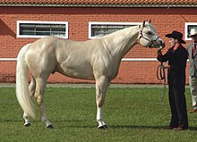
This horse, being built "downhill," will find it harder to shift the weight to the hind end.
A horse's "motor" is located in his hindquarters, and a horse that is heavy on the forehand (weight primarily on the forehand) is not able to properly move forward with impulsion. For good impulsion, a horse must either be balanced or have most of its weight tilted back toward its hindquarters.
Good riding aims to help transfer some of the animal's body weight back, getting the horse "off the forehand," but some riding disciplines require a greater amount of this transfer of weight (or "collection") than others. Sports such as dressage and show jumping require some of the greatest transfers of weight, while others, such as western pleasure, require a great deal less. However, it is beneficial for all horses to not travel "on the forehand," as this decreases the concussion placed on the front legs and their joints, thereby decreasing the risk of concussion-related lamenesses such as sidebone, ringbone, and others.
Certain conformational faults will encourage a horse to travel on the forehand, thereby making it more difficult for a rider to attain the shift in weight (although talented riders can usually train any horse to travel better with enough time). Horses that are built "downhill," with their hindquarters especially high, will be harder to collect.
The masseter, or cheek muscle, opens and closes the jaw and allows chewing. The Brachiocephalicus is a wide strap-like muscle that begins at the base of the skull behind the jaw and ends below the point of the shoulder to the humerus. This muscle moves the head from side to side, pulls the scapula forward, raises it in collection, and swings the foreleg forward. It is well developed for good movement, and too strong a rein contact inhibits free forward movement. The Sternocephalic begins at the jowl and ends at the sternum. This muscle moves the head and neck. In ewe-necked or bull-necked horses, this muscle is overdeveloped, and is difficult to get into a relaxed shape. The Rhomboideus begins at the Nuchal ligament and ends at the scapula. It lifts the shoulder and forehand, and pulls the scapula forward. The Splenius begins behind the poll and ends at the beginning of the Trapezius. This muscle turns and extends the neck, and makes up the topline if well developed. The Trapezius is a flat, sheet-like muscle that begins at the Occipital bone and ends at the spines of the 7th cervical and all the thoracic vertebrae. It lifts the shoulder and forehand, and if this muscle is well developed, the horse will work in a good outline. The Nuchal ligament begins at the poll and ends at the withers, and helps the muscles in the neck support the head. The deltoid begins at the scapula and ends at the humerus. The deltoid flexes the shoulder joint, and will load the shoulder if overdeveloped. The Supraspinatus begins below the trapezius and ends at the point of the shoulder; it maintains the shoulder in extension. The Latissimus dorsi begins at the lower thoracic vertebrae and ends at the back of the humerus; it flexes the shoulder and pulls the foreleg back. The Triceps flex the shoulder and extend the elbow; the Biceps flex the elbow and extend the shoulder. The pectoral muscles help pull the foreleg forward. The Triceps Brachii extend the elbow joint; the Biceps Brachii flex the elbow joint. The Extensor carpus extends the knee. The Flexor carpus flexes the knee. The Digital extensor extends the toe and knee. The digital flexor flexes the toe and knee and extends the elbow.
In human anatomy, the arm refers to the upper limb in common usage, although academically the term specifically means the upper arm between the glenohumeral joint and the elbow joint. The distal part of the upper limb between the elbow and the radiocarpal joint is known as the forearm or "lower" arm, and the extremity beyond the wrist is the hand.
The scapula, also known as the shoulder blade, is the bone that connects the humerus with the clavicle. Like their connected bones, the scapulae are paired, with each scapula on either side of the body being roughly a mirror image of the other. The name derives from the Classical Latin word for trowel or small shovel, which it was thought to resemble.
The humerus is a long bone in the arm that runs from the shoulder to the elbow. It connects the scapula and the two bones of the lower arm, the radius and ulna, and consists of three sections. The humeral upper extremity consists of a rounded head, a narrow neck, and two short processes. The body is cylindrical in its upper portion, and more prismatic below. The lower extremity consists of 2 epicondyles, 2 processes, and 3 fossae. As well as its true anatomical neck, the constriction below the greater and lesser tubercles of the humerus is referred to as its surgical neck due to its tendency to fracture, thus often becoming the focus of surgeons.
The biceps or biceps brachii are a large muscle that lies on the front of the upper arm between the shoulder and the elbow. Both heads of the muscle arise on the scapula and join to form a single muscle belly which is attached to the upper forearm. While the biceps crosses both the shoulder and elbow joints, its main function is at the elbow where it flexes the forearm and supinates the forearm. Both these movements are used when opening a bottle with a corkscrew: first biceps screws in the cork (supination), then it pulls the cork out (flexion).
The latissimus dorsi is a large, flat muscle on the back that stretches to the sides, behind the arm, and is partly covered by the trapezius on the back near the midline. The word latissimus dorsi comes from Latin and means "broadest [muscle] of the back", from "latissimus" and "dorsum". The pair of muscles are commonly known as "lats", especially among bodybuilders. The latissimus dorsi is the largest muscle in the upper body.
The axillary nerve or the circumflex nerve is a nerve of the human body, that originates from the brachial plexus at the level of the axilla (armpit) and carries nerve fibers from C5 and C6. The axillary nerve travels through the quadrangular space with the posterior circumflex humeral artery and vein to innervate the deltoid and teres minor.
The bench press, or chest press, is a weight training exercise where a person presses a weight upwards while lying horizontally on a weight training bench. Although the bench press is a compound movement, the muscles primarily used are the pectoralis major, the anterior deltoids, and the triceps, among other stabilizing muscles. A barbell is generally used to hold the weight, but a pair of dumbbells can also be used.
The upper limbs or upper extremities are the forelimbs of an upright-postured tetrapod vertebrate, extending from the scapulae and clavicles down to and including the digits, including all the musculatures and ligaments involved with the shoulder, elbow, wrist and knuckle joints. In humans, each upper limb is divided into the arm, forearm and hand, and is primarily used for climbing, lifting and manipulating objects.
The triceps, or triceps brachii, is a large muscle on the back of the upper limb of many vertebrates. It consists of 3 parts: the medial, lateral, and long head. It is the muscle principally responsible for extension of the elbow joint.
Equine conformation evaluates a horse's bone structure, musculature, and its body proportions in relation to each other. Undesirable conformation can limit the ability to perform a specific task. Although there are several faults with universal disadvantages, a horse's conformation is usually judged by what its intended use may be. Thus "form to function" is one of the first set of traits considered in judging conformation. A horse with poor form for a Grand Prix show jumper could have excellent conformation for a World Champion cutting horse, or to be a champion draft horse. Every horse has good and bad points of its conformation and many horses excel even with conformation faults.
The shoulder joint is structurally classified as a synovial ball-and-socket joint and functionally as a diarthrosis and multiaxial joint. It involves an articulation between the glenoid fossa of the scapula and the head of the humerus. Due to the very loose joint capsule that gives a limited interface of the humerus and scapula, it is the most mobile joint of the human body.
Dog anatomy comprises the anatomical study of the visible parts of the body of a domestic dog. Details of structures vary tremendously from breed to breed, more than in any other animal species, wild or domesticated, as dogs are highly variable in height and weight. The smallest known adult dog was a Yorkshire Terrier that stood only 6.3 cm (2.5 in) at the shoulder, 9.5 cm (3.7 in) in length along the head and body, and weighed only 113 grams (4.0 oz). The heaviest dog was an English Mastiff named Zorba, which weighed 314 pounds (142 kg). The tallest known adult dog is a Great Dane that stands 106.7 cm (42.0 in) at the shoulder.
The pull-down exercise is a strength training exercise designed to develop the latissimus dorsi muscle. It performs the functions of downward rotation and depression of the scapulae combined with adduction and extension of the shoulder joint.
The fascial compartments of arm refers to the specific anatomical term of the compartments within the upper segment of the upper limb of the body. The upper limb is divided into two segments, the arm and the forearm. Each of these segments is further divided into two compartments which are formed by deep fascia – tough connective tissue septa (walls). Each compartment encloses specific muscles and nerves.
The rear delt raise, also known as the rear deltoid raise, or rear shoulder raise is an exercise in weight training. This exercise is an isolation exercise that heavily works the posterior deltoid muscle. The movement is primarily limited to the two shoulder joints: the glenohumeral joint and the scapulothoracic joint. Scapular movement will also cause movement in the sternoclavicular joint and acromioclavicular joint. If the elbow bends during the extension exercises, it gravitates into a rowing motion.
The limbs of the horse are structures made of dozens of bones, joints, muscles, tendons, and ligaments that support the weight of the equine body. They include two apparatuses: the suspensory apparatus, which carries much of the weight, prevents overextension of the joint and absorbs shock, and the stay apparatus, which locks major joints in the limbs, allowing horses to remain standing while relaxed or asleep. The limbs play a major part in the movement of the horse, with the legs performing the functions of absorbing impact, bearing weight, and providing thrust. In general, the majority of the weight is borne by the front legs, while the rear legs provide propulsion. The hooves are also important structures, providing support, traction and shock absorption, and containing structures that provide blood flow through the lower leg. As the horse developed as a cursorial animal, with a primary defense mechanism of running over hard ground, its legs evolved to the long, sturdy, light-weight, one-toed form seen today.
The stay apparatus is an arrangement of muscles, tendons and ligaments that work together so that an animal can remain standing with virtually no muscular effort. It is best known as the mechanism by which horses can enter a light sleep while still standing up. The effect is that an animal can distribute its weight on three limbs while resting a fourth in a flexed, non-weight bearing position. The animal can periodically shift its weight to rest a different leg and thus all limbs are able to be individually rested, reducing overall wear and tear. The relatively slim legs of certain large mammals such as horses and cows would be subject to dangerous levels of fatigue if not for the stay apparatus.
This page is based on this Wikipedia article Text is available under the CC BY-SA 4.0 license; additional terms may apply. Images, videos and audio are available under their respective licenses.