Genetics of Down syndrome

Last updated
Chromosome 21 from Human Genome Program Human chromosome 21 description.png
Chromosome 21 from Human Genome Program
Normal human Karyotype. Karyotype.png
Normal human Karyotype.

Down syndrome is a chromosomal abnormality characterized by the presence of an extra copy of genetic material on chromosome 21, either in whole (trisomy 21) or part (such as due to translocations). The effects of the extra copy varies greatly from individual to individual, depending on the extent of the extra copy, genetic background, environmental factors, and random chance. Down syndrome can occur in all human populations, and analogous effects have been found in other species, such as chimpanzees and mice. In 2005, researchers have been able to create transgenic mice with most of human chromosome 21 (in addition to their normal chromosomes). [1]

Contents

A typical human karyotype is shown here. Every chromosome has two copies. In the bottom right, there are chromosomal differences between males (XY) and females (XX), which do not concern us. A typical human karyotype is designated as 46,XX or 46,XY, indicating 46 chromosomes with an XX arrangement for females and 46 chromosomes with an XY arrangement for males. [2] For this article, we will use females for the karyotype designation (46,XX).

Trisomy 21

Karyotype for trisomy Down syndrome. Notice the three copies of chromosome 21. Down Syndrome Karyotype.png
Karyotype for trisomy Down syndrome. Notice the three copies of chromosome 21.

Trisomy 21 (47,XY,+21) is caused by a meiotic nondisjunction event. [3] A typical gamete (either egg or sperm) has one copy of each chromosome (23 total). When it is combined with a gamete from the other parent during conception, the child has 46 chromosomes. However, with nondisjunction, a gamete is produced with an extra copy of chromosome 21 (the gamete has 24 chromosomes). When combined with a typical gamete from the other parent, the child now has 47 chromosomes, with three copies of chromosome 21. The trisomy 21 karyotype figure shows the chromosomal arrangement, with the prominent extra chromosome 21.

Trisomy 21 is the cause of approximately 95% of observed Down syndrome, with 88% coming from nondisjunction in the maternal gamete and 8% coming from nondisjunction in the paternal gamete. [4] Mitotic nondisjunction after conception would lead to mosaicism, and is discussed later.

Some cases have been reported of people with Down syndrome having children with trisomy 21. [5] In these cases (all from mothers), the ovaries were trisomy 21, leading to a secondary nondisjunction during gametogenesis and a gamete with an extra chromosome 21. Such Down syndrome trisomies are indistinguishable from Down syndrome trisomy created through meiotic nondisjunction.

Translocation

Balanced translocation with chromosomes 14 and 21q. Balanced Translocation.png
Balanced translocation with chromosomes 14 and 21q.
Translocation karyotype for Down syndrome with 14/21 Robertsonian translocation. Notice the three copies of 21q (two chromosomes 21 and the long arm of chromosome 21 fused to the short arm of a chromosome 14). Robertsonian translocation rob(14;21).png
Translocation karyotype for Down syndrome with 14/21 Robertsonian translocation. Notice the three copies of 21q (two chromosomes 21 and the long arm of chromosome 21 fused to the short arm of a chromosome 14).

The extra chromosome 21 material that causes Down syndrome may be due to a Robertsonian translocation. The long arm of chromosome 21 is attached to the long arm of another chromosome, often chromosome 14 [46,XX,t(14;21)] or itself [called an isochromosome, 45,XX,t(21;21)(q10;q10)] as seen in the translocation karyotype figure.

Translocation Down syndrome can be de novo; that is, not inherited but occurring at the time of an individual's conception, or may be inherited from a parent with a balanced translocation. The balanced translocation figure shows a 14/21 translocation between the long arms of chromosomes 14 and 21, where the other chromosomes are not shown. The individual has two copies of everything on chromosome 14, and two copies of all of the material on the long arm of chromosome 21 (21q). The derivative chromosome 21, which contains only heterochromatin, is lost; thus, the individual only has one copy of the material on the short arm of chromosome 21 (21p), but this appears to have no discernible effect. Individuals with this chromosomal arrangement have 45 chromosomes and are phenotypically normal. During meiosis, the chromosomal arrangement interferes with normal separation of chromosomes. Possible gametic arrangements are (see translocation karyotype figure):

Offspring from one parent with a balanced translocation and the other parent who has normal chromosomes. Translocation Down syndrome.png
Offspring from one parent with a balanced translocation and the other parent who has normal chromosomes.
1. translocated 14/21 and normal 14;
2. normal 14 and normal 21;
3. translocated 14/21 and normal 21;
4. normal 21 only;
5. normal 14 only;
6. translocated 14/21 only.

When combined with a normal gamete from the other parent, the first, fourth and fifth are lethal, leading to spontaneous abortion. The second, combined with a normal gamete from the other parent, gives rise to a typical child. The third leads to a translocation Down syndrome child. The last becomes a translocation carrier, like the parent.

Translocation Down syndrome is often referred to as familial Down syndrome. It is the cause of about 4.5% of the observed Down syndromes. [4] It does not show the maternal age effect, and is just as likely to have come from fathers as mothers.

Mosaicism

Mosaic Down syndrome is when some of the cells in the body do not have trisomy 21 and some cells have trisomy 21, an arrangement called a mosaic (46,XX/47,XX,+21). [6] [7] This can occur in one of two ways:

  • A nondisjunction event during an early cell division leads to a fraction of the cells with trisomy 21;
  • An anaphase lag of a chromosome 21 in a Down syndrome embryo leads to a fraction of euploid cells (2n cells), phenomenon described as "aneuploidy rescue".

There is considerable variability in the fraction of cells with trisomy 21, both as a whole and tissue-by-tissue. This is the cause of 1–2% of the observed Down syndromes. [4] There is evidence that mosaic Down syndrome may produce less developmental delay, on average, than full trisomy 21. [8]

The following diagram shows two possible mechanisms leading to mosaic trisomy 21.

           2n                    /    \               2n+1    2n-1
          2n+1                    /    \-> 1         /      \             2n+1      2n
Schematic of nondisjunction in mitosis.

Chromosomes in diploid somatic cell (2n). The nondisjunction leads to a daughter cell with a supernumerary chromosome (2n+1) and another cell missing a chromosome (2n-1).

Schematic of anaphase lag in mitosis.

Chromosomes in trisomy somatic cell (2n+1). One of the daughter cell is trisomic. The other daughter undergo anaphase lag: the supernumerary chromosome has a delayed movement during anaphase and fails to be included in the nucleusis. This mitotic segregation error restores the normal chromosome complement (2n).

Duplication of a portion of chromosome 21

Rarely, a region of chromosome 21 will undergo a duplication event. This will lead to extra copies of some, but not all, of the genes on chromosome 21 (46,XX,dup(21q)). If the duplicated region has genes that are responsible for Down syndrome physical and mental characteristics, such individuals will show those characteristics. This cause is very rare and no rate estimates are possible.

See also

Related Research Articles

<span class="mw-page-title-main">Chromosome</span> DNA molecule containing genetic material of a cell

A chromosome is a long DNA molecule with part or all of the genetic material of an organism. In most chromosomes the very long thin DNA fibers are coated with packaging proteins; in eukaryotic cells the most important of these proteins are the histones. These proteins, aided by chaperone proteins, bind to and condense the DNA molecule to maintain its integrity. These chromosomes display a complex three-dimensional structure, which plays a significant role in transcriptional regulation.

<span class="mw-page-title-main">Trisomy</span> Abnormal presence of three copies of a particular chromosome

A trisomy is a type of polysomy in which there are three instances of a particular chromosome, instead of the normal two. A trisomy is a type of aneuploidy.

<span class="mw-page-title-main">Karyotype</span> Photographic display of total chromosome complement in a cell

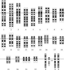
A karyotype is the general appearance of the complete set of chromosomes in the cells of a species or in an individual organism, mainly including their sizes, numbers, and shapes. Karyotyping is the process by which a karyotype is discerned by determining the chromosome complement of an individual, including the number of chromosomes and any abnormalities.

<span class="mw-page-title-main">Aneuploidy</span> Presence of an abnormal number of chromosomes in a cell

Aneuploidy is the presence of an abnormal number of chromosomes in a cell, for example a human cell having 45 or 47 chromosomes instead of the usual 46. It does not include a difference of one or more complete sets of chromosomes. A cell with any number of complete chromosome sets is called a euploid cell.

<span class="mw-page-title-main">Cytogenetics</span> Branch of genetics

Cytogenetics is essentially a branch of genetics, but is also a part of cell biology/cytology, that is concerned with how the chromosomes relate to cell behaviour, particularly to their behaviour during mitosis and meiosis. Techniques used include karyotyping, analysis of G-banded chromosomes, other cytogenetic banding techniques, as well as molecular cytogenetics such as fluorescence in situ hybridization (FISH) and comparative genomic hybridization (CGH).

<span class="mw-page-title-main">Patau syndrome</span> Chromosomal disorder in which there are three copies of chromosome 13

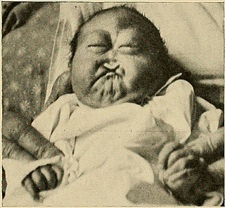
Patau syndrome is a syndrome caused by a chromosomal abnormality, in which some or all of the cells of the body contain extra genetic material from chromosome 13. The extra genetic material disrupts normal development, causing multiple and complex organ defects.

<span class="mw-page-title-main">Trisomy 18</span> Chromosomal disorder in which there are three copies of chromosome 18

Trisomy 18, also known as Edwards syndrome, is a genetic disorder caused by the presence of a third copy of all or part of chromosome 18. Many parts of the body are affected. Babies are often born small and have heart defects. Other features include a small head, small jaw, clenched fists with overlapping fingers, and severe intellectual disability.

<span class="mw-page-title-main">Nondisjunction</span> Failure to separate properly during cell division

Nondisjunction is the failure of homologous chromosomes or sister chromatids to separate properly during cell division (mitosis/meiosis). There are three forms of nondisjunction: failure of a pair of homologous chromosomes to separate in meiosis I, failure of sister chromatids to separate during meiosis II, and failure of sister chromatids to separate during mitosis. Nondisjunction results in daughter cells with abnormal chromosome numbers (aneuploidy).

<span class="mw-page-title-main">Chromosomal translocation</span> Phenomenon that results in unusual rearrangement of chromosomes

In genetics, chromosome translocation is a phenomenon that results in unusual rearrangement of chromosomes. This includes balanced and unbalanced translocation, with two main types: reciprocal, and Robertsonian translocation. Reciprocal translocation is a chromosome abnormality caused by exchange of parts between non-homologous chromosomes. Two detached fragments of two different chromosomes are switched. Robertsonian translocation occurs when two non-homologous chromosomes get attached, meaning that given two healthy pairs of chromosomes, one of each pair "sticks" and blends together homogeneously.

<span class="mw-page-title-main">Mosaic (genetics)</span> Condition in multi-cellular organisms

Mosaicism or genetic mosaicism is a condition in which a multicellular organism possesses more than one genetic line as the result of genetic mutation. This means that various genetic lines resulted from a single fertilized egg. Mosaicism is one of several possible causes of chimerism, wherein a single organism is composed of cells with more than one distinct genotype.

<span class="mw-page-title-main">XXYY syndrome</span> Extra X and Y chromosome in males

XXYY syndrome is a sex chromosome anomaly in which males have 2 extra chromosomes, one X and one Y chromosome. Human cells usually contain two sex chromosomes, one from the mother and one from the father. Usually, females have two X chromosomes (XX) and males have one X and one Y chromosome (XY). The appearance of at least one Y chromosome with a properly functioning SRY gene makes a male. Therefore, humans with XXYY are genotypically male. Males with XXYY syndrome have 48 chromosomes instead of the typical 46. This is why XXYY syndrome is sometimes written as 48, XXYY syndrome or 48, XXYY. It affects an estimated one in every 18,000–40,000 male births.

<span class="mw-page-title-main">Gonadal dysgenesis</span> Congenital disorder of the reproductive system

Gonadal dysgenesis is classified as any congenital developmental disorder of the reproductive system in humans. It is atypical development of gonads in an embryo,. One type of gonadal dysgenesis is the development of functionless, fibrous tissue, termed streak gonads, instead of reproductive tissue. Streak gonads are a form of aplasia, resulting in hormonal failure that manifests as sexual infantism and infertility, with no initiation of puberty and secondary sex characteristics.

A chromosomal abnormality, chromosomal anomaly, chromosomal aberration, chromosomal mutation, or chromosomal disorder, is a missing, extra, or irregular portion of chromosomal DNA. These can occur in the form of numerical abnormalities, where there is an atypical number of chromosomes, or as structural abnormalities, where one or more individual chromosomes are altered. Chromosome mutation was formerly used in a strict sense to mean a change in a chromosomal segment, involving more than one gene. Chromosome anomalies usually occur when there is an error in cell division following meiosis or mitosis. Chromosome abnormalities may be detected or confirmed by comparing an individual's karyotype, or full set of chromosomes, to a typical karyotype for the species via genetic testing.

Emanuel syndrome, also known as derivative 22 syndrome, or der(22) syndrome, is a rare disorder associated with multiple congenital anomalies, including profound intellectual disability, preauricular skin tags or pits, and conotruncal heart defects. It can occur in offspring of carriers of the constitutional chromosomal translocation t(11;22)(q23;q11), owing to a 3:1 meiotic malsegregation event resulting in partial trisomy of chromosomes 11 and 22. An unbalanced translocation between chromosomes 11 and 22 is described as Emanuel syndrome. It was first described in 1980 by American medical researchers Beverly S. Emanuel and Elaine H. Zackai, and a consortium of European scientists the same year.

<span class="mw-page-title-main">Trisomy 22</span> Medical condition

Trisomy 22 is a chromosomal disorder in which three copies of chromosome 22 are present rather than two. It is a frequent cause of spontaneous abortion during the first trimester of pregnancy. Progression to the second trimester and live birth are rare. This disorder is found in individuals with an extra copy or a variation of chromosome 22 in some or all cells of their bodies.

<span class="mw-page-title-main">Klinefelter syndrome</span> Human chromosomal condition

Klinefelter syndrome (KS), also known as 47,XXY, is an aneuploid genetic condition where a male has an additional copy of the X chromosome. The primary features are infertility and small, poorly functioning testicles. Usually, symptoms are subtle and subjects do not realize they are affected. Sometimes, symptoms are more evident and may include weaker muscles, greater height, poor motor coordination, less body hair, breast growth, and less interest in sex. Often, these symptoms are noticed only at puberty. Intelligence is usually average, but reading difficulties and problems with speech are more common.

45,X/46,XY mosaicism, also known as X0/XY mosaicism and mixed gonadal dysgenesis, is a mutation of sex development in humans associated with sex chromosome aneuploidy and mosaicism of the Y chromosome. This is called a mosaic karyotype because, like tiles in mosaic floors or walls, there is more than one type of cell. It is a fairly rare chromosomal disorder at birth, with an estimated incidence rate of about 1 in 15,000 live births. Mosaic loss of the Y chromosome in previously non-mosaic men grows increasingly common with age.

<span class="mw-page-title-main">Tetrasomy X</span> Chromosomal disorder with 4 X chromosomes

Tetrasomy X, also known as 48,XXXX, is a chromosomal disorder in which a female has four, rather than two, copies of the X chromosome. It is associated with intellectual disability of varying severity, characteristic "coarse" facial features, heart defects, and skeletal anomalies such as increased height, clinodactyly, and radioulnar synostosis. Tetrasomy X is a rare condition, with few medically recognized cases; it is estimated to occur in approximately 1 in 50,000 females.

<span class="mw-page-title-main">Pentasomy X</span> Chromosomal disorder

Pentasomy X, also known as 49,XXXXX, is a chromosomal disorder in which a female has five, rather than two, copies of the X chromosome. Pentasomy X is associated with short stature, intellectual disability, characteristic facial features, heart defects, skeletal anomalies, and pubertal and reproductive abnormalities. The condition is exceptionally rare, with an estimated prevalence between 1 in 85,000 and 1 in 250,000.

<span class="mw-page-title-main">Trisomy X</span> Chromosome disorder in women

Trisomy X, also known as triple X syndrome and characterized by the karyotype 47,XXX, is a chromosome disorder in which a female has an extra copy of the X chromosome. It is relatively common and occurs in 1 in 1,000 females but it is rarely diagnosed; fewer than 10 per cent of those with the condition know they have it.

References

  1. BBC News (22 September 2005). "Down's syndrome recreated in mice" . Retrieved 2006-06-14.
  2. For a description of human karyotype see Mitelman, F. (1995). ISCN 1995 : an international system for human cytogenetic nomenclature (1995) : recommendations of the International Standing Committee on Human Cytogenetic Nomenclature, Memphis, Tennessee, USA, October 9-13, 1994.
  3. There is a nice animation that shows nondisjunction at "Meiotic nondisjunction animation". Archived from the original on 2006-05-17. Retrieved 2006-07-01.
  4. 1 2 3 "Down syndrome occurrence rates (NIH)". Archived from the original on 2006-05-17. Retrieved 2006-06-02.
  5. For an example of mother with mosaic Down syndrome, see Karkany, J. (1971). Congenital Malformations. Chicago: Year Book Medical Publishers, Inc. pp. 319–322. ISBN   0-8151-9098-0.
  6. Mosaic Down Syndrome on the Web
  7. International Mosaic Down Syndrome Association
  8. Leshin, L. (2000). "Mosaic Down Syndrome" . Retrieved 2006-06-02.