Glaucoma valve

Last updated
Flow of the aqueous humour Flow of aqueous humour eye EDA02.JPG
Flow of the aqueous humour

A glaucoma valve is a medical shunt used in the treatment of glaucoma to reduce the eye's intraocular pressure (IOP).

Contents

Mechanism

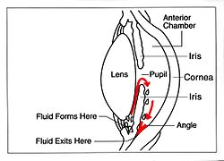
The device works by bypassing the trabecular meshwork and redirecting the outflow of aqueous humour through a small tube into an outlet chamber or bleb. The IOP generally decreases from around 33 to 10 mmHg by removing aqueous on average 2.75 microliters/min. [1]

Types

The first glaucoma drainage implant was developed in 1966. [2] Following on the success of the Molteno implant, several varieties of device have been developed from the original, the Baerveldt tube shunt, or the valved implants, such as the Ahmed glaucoma valve implant and the later generation pressure ridge Molteno implants. These are indicated for glaucoma patients not responding to maximal medical therapy, with previous failed guarded filtering surgery (trabeculectomy). The flow tube is inserted into the anterior chamber of the eye and the plate is implanted underneath the conjunctiva to allow flow of aqueous fluid out of the eye into a chamber called a bleb.

The ExPress Mini Shunt is a newer, non-valved device that was originally designed to provide a direct conduit from the anterior chamber to the sub-conjunctival space or bleb. In this position it was unstable and tended to erode through the conjunctiva. Now the more common use is as a modification of the trabeculectomy procedure, placed under a scleral flap, replacing the sclerostomy step (see trabeculectomy).

In comparison to the glaucoma drainage devices that use an ab externo procedure, ab interno implants, such as the Xen Gel Stent, are transscleral implants to channel aqueous humor into the non-dissected Tenon's space, creating a subconjunctival drainage area similar to a bleb. [3] [4] The implants are transscleral and different from more other ab interno implants that do not create a transscleral drainage, such as iStent, CyPass, or Hydrus. [5]

A Cochrane Review of various aqueous shunts and modifications found that the Baerveldt implant may result in lower IOP than the Ahmed implant, but it was unclear if the difference in IOP reduction was clinically significant. [6] The review suggests that practitioners should operate with the devices that they are most comfortable with, and have the most experience using.

Indications

The glaucoma valve implant is indicated for glaucoma patients not responding to maximal medical therapy, with previous failed guarded filtering surgery (trabeculectomy) or in cases where conventional drainage surgery is unlikely to succeed. Common situations where the use of a glaucoma implant as a primary procedure is indicated include

Surgical technique

The flow tube is inserted into the anterior chamber of the eye and the plate is implanted underneath the conjunctiva to allow flow of aqueous fluid out of the eye.

Complications

The majority of complications occur shortly after surgery. These are generally related to high pressure (due to inflammation following surgery) or low pressure (too much aqueous flow through the tube). Periods of low pressure which are more associated with non-valved shunts, can cause retinal detachments, hypotony maculopathy or haemorrhages. Periods of high pressures, which are more associated with valved shunts, are detrimental to the optic nerve. Long term complications of this surgery include diplopia and corneal oedema.

There are also device related complications, which will require surgical revision. For example, erosion, where the conjunctiva erodes over the shunt leaving it exposed, the condition of which may be revised or prevented in advance by the use of amniotic membrane, [12] The ologen collagen matrix facilitates tissue regeneration and its application over the site of device implantation can strengthen tissue recovery, reducing possibility of erosion. [13] [14] [12] [15] [16]

When the device malfunctions it may need to be replaced. Possible scenarios include blockage, where a particle becomes logged in the tube line blocking flow; retraction, where the tube line slips out of correct position such that flow is inhibited or halted; valve failure, where the valve stops working blocking flow completely.

Surgical failure occurs due to the ongoing scarring over the conjunctival dissipation segment of the shunt may become too thick for the aqueous humor to filter through. This may require preventive measures using anti-fibrotic medication like 5-fluorouracil (5FU) or Mitomycin-C (during the procedure), or creating a necessity for revision surgery with the sole or combinative use of biodegradable spacer or collagen matrix implant. [6] A Cochrane Review comparing aqueous shunt surgery with and without Mitomycin-C did not find benefit or harm associated with the intervention. [17]

See also

Related Research Articles

<span class="mw-page-title-main">Glaucoma</span> Group of eye diseases

Glaucoma is a group of eye diseases that lead to damage of the optic nerve, which transmits visual information from the eye to the brain. Glaucoma may cause vision loss if left untreated. It has been called the "silent thief of sight" because the loss of vision usually occurs slowly over a long period of time. A major risk factor for glaucoma is increased pressure within the eye, known as intraocular pressure (IOP). It is associated with old age, a family history of glaucoma, and certain medical conditions or medications. The word glaucoma comes from the Ancient Greek word γλαυκός, meaning 'gleaming, blue-green, gray'.

<span class="mw-page-title-main">Eye surgery</span> Surgery performed on the eye or its adnexa

Eye surgery, also known as ophthalmic surgery or ocular surgery, is surgery performed on the eye or its adnexa. Eye surgery is part of ophthalmology and is performed by an ophthalmologist or eye surgeon. The eye is a fragile organ, and requires due care before, during, and after a surgical procedure to minimize or prevent further damage. An eye surgeon is responsible for selecting the appropriate surgical procedure for the patient, and for taking the necessary safety precautions. Mentions of eye surgery can be found in several ancient texts dating back as early as 1800 BC, with cataract treatment starting in the fifth century BC. It continues to be a widely practiced class of surgery, with various techniques having been developed for treating eye problems.

<span class="mw-page-title-main">Intraocular pressure</span> Fluid pressure inside the eye

Intraocular pressure (IOP) is the fluid pressure inside the eye. Tonometry is the method eye care professionals use to determine this. IOP is an important aspect in the evaluation of patients at risk of glaucoma. Most tonometers are calibrated to measure pressure in millimeters of mercury (mmHg).

<span class="mw-page-title-main">Trabecular meshwork</span> Area of tissue in the eye

The trabecular meshwork is an area of tissue in the eye located around the base of the cornea, near the ciliary body, and is responsible for draining the aqueous humor from the eye via the anterior chamber.

George Baerveldt, M.B.Ch.B., was a Professor in the Department of Ophthalmology at the University of California, Irvine.

<span class="mw-page-title-main">Trabeculectomy</span> Surgical removal of trabecular tissue in the eye

Trabeculectomy is a surgical procedure used in the treatment of glaucoma to relieve intraocular pressure by removing part of the eye's trabecular meshwork and adjacent structures. It is the most common glaucoma surgery performed and allows drainage of aqueous humor from within the eye to underneath the conjunctiva where it is absorbed. This outpatient procedure was most commonly performed under monitored anesthesia care using a retrobulbar block or peribulbar block or a combination of topical and subtenon anesthesia. Due to the higher risks associated with bulbar blocks, topical analgesia with mild sedation is becoming more common. Rarely general anesthesia will be used, in patients with an inability to cooperate during surgery.

<span class="mw-page-title-main">Glaucoma surgery</span> Type of eye surgery

Glaucoma is a group of diseases affecting the optic nerve that results in vision loss and is frequently characterized by raised intraocular pressure (IOP). There are many glaucoma surgeries, and variations or combinations of those surgeries, that facilitate the escape of excess aqueous humor from the eye to lower intraocular pressure, and a few that lower IOP by decreasing the production of aqueous humor.

<span class="mw-page-title-main">Pterygium</span> Winglike triangular membrane

A pterygium is any wing-like triangular membrane occurring in the neck, eyes, knees, elbows, ankles or digits.

In medicine, the term cheesewiring or cheesewire effect describes any process in which cells or intercellular matrix are dissected or extruded either by the material being pressed through a taut element, or by the tension of a taut element pulling through tissue. The procedure is typically conducted in a surgical setting.

<span class="mw-page-title-main">Canine glaucoma</span> Optic nerve disease in dogs

Canine glaucoma refers to a group of diseases in dogs that affect the optic nerve and involve a loss of retinal ganglion cells in a characteristic pattern. An intraocular pressure greater than 22 mmHg (2.9 kPa) is a significant risk factor for the development of glaucoma. Untreated glaucoma in dogs leads to permanent damage of the optic nerve and resultant visual field loss, which can progress to blindness.

<span class="mw-page-title-main">Boston keratoprosthesis</span> Prosthetic cornea

Boston keratoprosthesis is a collar button design keratoprosthesis or artificial cornea. It is composed of a front plate with a stem, which houses the optical portion of the device, a back plate and a titanium locking c-ring. It is available in type I and type II formats. The type I design is used much more frequently than the type II which is reserved for severe end stage dry eye conditions and is similar to the type I except it has a 2 mm anterior nub designed to penetrate through a tarsorrhaphy. The type I format will be discussed here as it is more commonly used.

<span class="mw-page-title-main">Bleb (medicine)</span> Blister-like protrusion filled with serous fluid

In medicine, a bleb is a blister-like protrusion filled with serous fluid. Blebs can form in a number of tissues by different pathologies, including frostbite and can "appear and disappear within a short time interval".

The Trabectome is a surgical device that can be used for ab interno trabeculotomy, a minimally invasive glaucoma surgery for the surgical management of adult, juvenile, and infantile glaucoma. The trabecular meshwork is a major site of resistance to aqueous humor outflow. As angle surgeries such as Trabectome follow the physiologic outflow pathway, the risk of complications is significantly lower than filtering surgeries. Hypotony with damage to the macula, can occur with pressures below 5 mmHg, for instance, after traditional trabeculectomy, because of the episcleral venous pressure limit. The Trabectome handpiece is inserted into the anterior chamber, its tip positioned into Schlemm's canal, and advanced to the left and to the right. Different from cautery, the tip generates plasma to molecularize the trabecular meshwork and remove it drag-free and with minimal thermal effect. Active irrigation of the trabectome surgery system helps to keep the anterior chamber formed during the procedure and precludes the need for ophthalmic viscoelastic devices. Viscoelastic devices tend to trap debris or gas bubbles and diminish visualization. The Trabectome decreases the intra-ocular pressure typically to a mid-teen range and reduces the patient's requirement to take glaucoma eye drops and glaucoma medications. The theoretically lowest pressure that can be achieved is equal to 8 mmHg in the episcleral veins. This procedure is performed through a small incision and can be done on an outpatient basis.

Micro-invasive glaucoma surgery (MIGS) is the latest advance in surgical treatment for glaucoma, which aims to reduce intraocular pressure by either increasing outflow of aqueous humor or reducing its production. MIGS comprises a group of surgical procedures which share common features. MIGS procedures involve a minimally invasive approach, often with small cuts or micro-incisions through the cornea that causes the least amount of trauma to surrounding scleral and conjunctival tissues. The techniques minimize tissue scarring, allowing for the possibility of traditional glaucoma procedures such as trabeculectomy or glaucoma valve implantation to be performed in the future if needed.

<span class="mw-page-title-main">Secondary glaucoma</span>

Secondary glaucoma is a collection of progressive optic nerve disorders associated with a rise in intraocular pressure (IOP) which results in the loss of vision. In clinical settings, it is defined as the occurrence of IOP above 21 mmHg requiring the prescription of IOP-managing drugs. It can be broadly divided into two subtypes: secondary open-angle glaucoma and secondary angle-closure glaucoma, depending on the closure of the angle between the cornea and the iris. Principal causes of secondary glaucoma include optic nerve trauma or damage, eye disease, surgery, neovascularization, tumours and use of steroid and sulfa drugs. Risk factors for secondary glaucoma include uveitis, cataract surgery and also intraocular tumours. Common treatments are designed according to the type and the underlying causative condition, in addition to the consequent rise in IOP. These include drug therapy, the use of miotics, surgery or laser therapy.

Uveitis–glaucoma–hyphaema (UGH) syndrome, also known as Ellingson syndrome, is a complication of cataract surgery, caused by intraocular lens subluxation or dislocation. The chafing of mispositioned intraocular lens over iris, ciliary body or iridocorneal angle cause elevated intraocular pressure (IOP) anterior uveitis and hyphema. It is most commonly caused by anterior chamber IOLs and sulcus IOLs but, the condition can be seen with any type of IOL, including posterior chamber lenses and cosmetic iris implants.

<span class="mw-page-title-main">Uveitic glaucoma</span> Glaucoma caused by uveitis or its treatments

Uveitic glaucoma is most commonly a progression stage of noninfectious anterior uveitis or iritis.

Ocular hypotony, or ocular hypotension, or shortly hypotony, is the medical condition in which intraocular pressure (IOP) of the eye is very low.

Hypotony maculopathy is maculopathy due to very low intraocular pressure known as ocular hypotony. Maculopathy occurs either due to increased outflow of aqueous humor through angle of anterior chamber or less commonly, due to decreased aqueous humor secretion by ciliary body.

References

  1. Brubaker, Richard F. (1991-12-01). "Flow of Aqueous Humor in Humans [The Friedenwald Lecture]". Investigative Ophthalmology & Visual Science. 32 (13): 3145–66. PMID   1748546.[ permanent dead link ]
  2. "Archived copy" (PDF). Archived from the original (PDF) on 2015-01-13. Retrieved 2012-05-15.{{cite web}}: CS1 maint: archived copy as title (link)[ full citation needed ]
  3. Lewis RA (Aug 2014). "Ab interno approach to the subconjunctival space using a collagen glaucoma stent". J Cataract Refract Surg. 40 (8): 1301–6. doi:10.1016/j.jcrs.2014.01.032. PMID   24943904.
  4. "Xen Gel Stent". AqueSys. Archived from the original on 29 June 2015. Retrieved 27 June 2015.
  5. "Advances in Glaucoma Filtration Surgery". Glaucoma Today. Retrieved 27 June 2015.
  6. 1 2 Tseng VL, Coleman AL, Chang MY, Caprioli J (2017). "Aqueous shunts for glaucoma". Cochrane Database Syst Rev. 2017 (7): CD004918. doi:10.1002/14651858.CD004918.pub3. PMC   5580949 . PMID   28750481.
  7. Oscar, Albis-Donado; Gil-Carrasco, Gil-Carrasco; Romero-Quijada, Romero-Quijada; Thomas, Thomas (2010). "Evaluation of ahmed glaucoma valve implantation through a needle-generated scleral tunnel in Mexican children with glaucoma". Indian Journal of Ophthalmology. 58 (5): 365–73. doi: 10.4103/0301-4738.67039 . PMC   2992909 . PMID   20689189.
  8. Molteno, AC; Polkinghorne, PJ; Bowbyes, JA (1986). "The vicryl tie technique for inserting a draining implant in the treatment of secondary glaucoma". Australian and New Zealand Journal of Ophthalmology. 14 (4): 343–54. doi: 10.1111/j.1442-9071.1986.tb00470.x . PMID   3814422.
  9. Ayyala, RS; Zurakowski, D; Monshizadeh, R; Hong, CH; Richards, D; Layden, WE; Hutchinson, BT; Bellows, AR (2002). "Comparison of double-plate Molteno and Ahmed glaucoma valve in patients with advanced uncontrolled glaucoma". Ophthalmic Surgery and Lasers. 33 (2): 94–101. doi:10.3928/1542-8877-20020301-04. PMID   11942556.
  10. Budenz, DL; Barton, K; Feuer, WJ; Schiffman, J; Costa, VP; Godfrey, DG; Buys, YM; Ahmed Baerveldt Comparison Study Group (2011). "Treatment outcomes in the Ahmed Baerveldt Comparison Study after 1 year of follow-up". Ophthalmology. 118 (3): 443–52. doi:10.1016/j.ophtha.2010.07.016. PMC   3020266 . PMID   20932583.
  11. Christakis, PG; Kalenak, JW; Zurakowski, D; Tsai, JC; Kammer, JA; Harasymowycz, PJ; Ahmed, II (2011). "The Ahmed Versus Baerveldt study: One-year treatment outcomes". Ophthalmology. 118 (11): 2180–9. doi:10.1016/j.ophtha.2011.05.004. PMID   21889801.
  12. 1 2 Rosentreter, A; Schield AM; Dinslage S; Dietlein TS (2012). "Biodegradable implant for tissue repair after glaucoma drainage device surgery". J Glaucoma. 21 (2): 76–8. doi:10.1097/IJG.0b013e3182027ab0. PMID   21278584. S2CID   40206358.
  13. Rho, S; Sung Y; Ma KT; Rho SH; Kim CY (2015). "Bleb Analysis and Short-Term Results of Biodegradable Collagen Matrix-Augmented Ahmed Glaucoma Valve Implantation: 6-Month Follow-up". Invest Ophthalmol Vis Sci. 56 (10): 5896–903. doi: 10.1167/iovs.15-17480 . PMID   26348639.
  14. Johnson, MS; Sarkisian SR Jr. (2014). "Using a collagen matrix implant (Ologen) versus mitomycin-C as a wound healing modulator in trabeculectomy with the Ex-PRESS mini glaucoma device: a 12-month retrospective review". J Glaucoma. 23 (9): 649–52. doi:10.1097/IJG.0000000000000018. PMID   24240882. S2CID   45334774.
  15. Rosentreter, A; Mellein AC; Konen WW; Dietlein TS (2010). "Capsule excision and Ologen™ implantation for revision after glaucoma drainage device surgery". Graefes Arch Clin Exp Ophthalmol. 248 (9): 1319–24. doi:10.1007/s00417-010-1385-y. PMID   20405139. S2CID   10384646.
  16. Aptel, F; Dumas S; Denis P (2009). "Ultrasound biomicroscopy and optical coherence tomography imaging of filtering blebs after deep sclerectomy with new collagen implant". Eur J Ophthalmol. 19 (2): 223–30. doi:10.1177/112067210901900208. PMID   19253238. S2CID   22594085.
  17. Foo VH, Htoon HM, Welsbie DS, Perera SA (2019). "Aqueous shunts with mitomycin C versus aqueous shunts alone for glaucoma". Cochrane Database Syst Rev. 2019 (4): CD011875. doi:10.1002/14651858.CD011875.pub2. PMC   6472957 . PMID   30999387.