Related Research Articles

A computed tomography scan, formerly called computed axial tomography scan, is a medical imaging technique used to obtain detailed internal images of the body. The personnel that perform CT scans are called radiographers or radiology technologists.

A bronchus is a passage or airway in the lower respiratory tract that conducts air into the lungs. The first or primary bronchi to branch from the trachea at the carina are the right main bronchus and the left main bronchus. These are the widest bronchi, and enter the right lung, and the left lung at each hilum. The main bronchi branch into narrower secondary bronchi or lobar bronchi, and these branch into narrower tertiary bronchi or segmental bronchi. Further divisions of the segmental bronchi are known as fourth order, fifth order, and sixth order segmental bronchi, or grouped together as subsegmental bronchi. The bronchi, when too narrow to be supported by cartilage, are known as bronchioles. No gas exchange takes place in the bronchi.

Scimitar syndrome, or congenital pulmonary venolobar syndrome, is a rare congenital heart defect characterized by anomalous venous return from the right lung. This anomalous pulmonary venous return can be either partial (PAPVR) or total (TAPVR). The syndrome associated with PAPVR is more commonly known as Scimitar syndrome after the curvilinear pattern created on a chest radiograph by the pulmonary veins that drain to the inferior vena cava. This radiographic density often has the shape of a scimitar, a type of curved sword. The syndrome was first described by Catherine Neill in 1960.
Allergic bronchopulmonary aspergillosis (ABPA) is a condition characterised by an exaggerated response of the immune system to the fungus Aspergillus. It occurs most often in people with asthma or cystic fibrosis. Aspergillus spores are ubiquitous in soil and are commonly found in the sputum of healthy individuals. A. fumigatus is responsible for a spectrum of lung diseases known as aspergilloses.
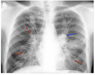
A bronchocele is a segment of bronchus that is filled with mucus and completely enclosed so the mucus cannot drain out. This segment is usually dilated. It is also referred to as bronchial mucocele. If there is no obstruction to the flow of mucus, it is called mucoid impaction of bronchus. A bronchocele results from obstruction of the bronchus. Overproduction of mucus can also contribute. Obstruction may occur due to scarring, a tumor, or congenital atresia.

In radiology, the deep sulcus sign on a supine chest radiograph is an indirect indicator of a pneumothorax. In a supine film, it appears as a deep, lucent, ipsilateral costophrenic angle within the nondependent portions of the pleural space as opposed to the apex when the patient is upright. The costophrenic angle is abnormally deepened when the pleural air collects laterally, producing the deep sulcus sign.
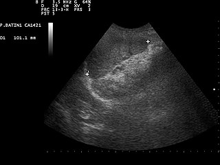
Focused assessment with sonography in trauma is a rapid bedside ultrasound examination performed by surgeons, emergency physicians, and paramedics as a screening test for blood around the heart or abdominal organs (hemoperitoneum) after trauma. There is also the extended FAST (eFAST) which includes some additional ultrasound views to assess for pneumothorax.

In radiology, the air crescent sign is a finding on chest radiograph and computed tomography that is crescenteric and radiolucent, due to a lung cavity that is filled with air and has a round radiopaque mass. Classically, it is due to an aspergilloma, a form of aspergillosis, that occurs when the fungus Aspergillus grows in a cavity in the lung. It is also referred as Monad sign.

Lung cancer screening refers to cancer screening strategies used to identify early lung cancers before they cause symptoms, at a point where they are more likely to be curable. Lung cancer screening is critically important because of the incidence and prevalence of lung cancer. More than 235,000 new cases of lung cancer are expected in the United States in 2021 with approximately 130,000 deaths expected in 2021. In addition, at the time of diagnosis, 57% of lung cancers are discovered in advanced stages, meaning they are more widespread or aggressive cancers. Because there is a substantially higher probability of long-term survival following treatment of localized (60%) versus advanced stage (6%) lung cancer, lung cancer screening aims to diagnose the disease in the localized stage.
In radiology, the tree-in-bud sign is a finding on a CT scan that indicates some degree of airway obstruction. The tree-in-bud sign is a nonspecific imaging finding that implies impaction within bronchioles, the smallest airway passages in the lung. The differential for this finding includes malignant and inflammatory etiologies, either infectious or sterile. This includes fungal infections, mycobacterial infections such as tuberculosis or mycobacterium avium intracellulare, bronchopneumonia, chronic aspiration pneumonia, cystic fibrosis or cellular impaction from bronchovascular spread of malignancy, as can occur with breast cancer, leukemia or lymphoma. It also includes lung manifestations of autoimmune diseases such as Sjögren syndrome or rheumatoid arthritis.

Diaphragmatic rupture is a tear of the diaphragm, the muscle across the bottom of the ribcage that plays a crucial role in breathing. Most commonly, acquired diaphragmatic tears result from physical trauma. Diaphragmatic rupture can result from blunt or penetrating trauma and occurs in about 0.5% of all people with trauma.

A lung nodule or pulmonary nodule is a relatively small focal density in the lung. A solitary pulmonary nodule (SPN) or coin lesion, is a mass in the lung smaller than three centimeters in diameter. A pulmonary micronodule has a diameter of less than three millimetres. There may also be multiple nodules.
In radiology, the Golden S sign, also known as the S sign of Golden, is a radiologic sign seen on chest X-ray that suggests a central lung mass or a lung collapse. It was first described by, and subsequently named after, Dr Ross Golden (1889–1975) in 1925 in association with bronchial carcinoma, but it is also seen in metastatic cancer, enlarged lymph nodes, and collapse of the right upper lobe of the lung.

Ground-glass opacity (GGO) is a finding seen on chest x-ray (radiograph) or computed tomography (CT) imaging of the lungs. It is typically defined as an area of hazy opacification (x-ray) or increased attenuation (CT) due to air displacement by fluid, airway collapse, fibrosis, or a neoplastic process. When a substance other than air fills an area of the lung it increases that area's density. On both x-ray and CT, this appears more grey or hazy as opposed to the normally dark-appearing lungs. Although it can sometimes be seen in normal lungs, common pathologic causes include infections, interstitial lung disease, and pulmonary edema.

In radiology, honeycombing or "honeycomb lung" is the radiological appearance seen with widespread pulmonary fibrosis and is defined by the presence of small cystic spaces with irregularly thickened walls composed of fibrous tissue. Dilated and thickened terminal and respiratory bronchioles produce cystic airspaces, giving a honeycomb appearance on chest x-rays. Honeycomb cysts often predominate in the peripheral and pleural/subpleural lung regions regardless of their cause.

Pneumatosis is the abnormal presence of air or other gas within tissues.

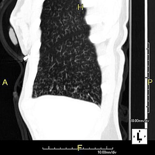
A focal lung pneumatosis is an enclosed pocket of air or gas in the lung and includes blebs, bullae, pulmonary cysts, and lung cavities. Blebs and bullae can be classified by their wall thickness.

Crazy paving refers to a pattern seen on computed tomography of the chest, involving lobular septal thickening with variable alveolar filling. The finding is seen in pulmonary alveolar proteinosis, and other diseases. Its name comes from its resemblance to irregular paving stones, called crazy pavings.
Juxtaphrenic peak sign is a radiographic sign seen in lobar collapse or after lobectomy of the lung. This sign was first described by Katten and colleagues in 1980, and therefore, it is also called Katten's sign. The juxtaphrenic peak is most commonly caused due to the traction from the inferior accessory fissure. The prevalence of the juxtaphrenic peak sign increases gradually during the weeks after lobectomy of the lung.
The cheerio sign is a radiologic sign that has been used to describe both pulmonary nodules and tears in the glenoid labrum, named after its resemblance to the popular breakfast cereal "Cheerios".
References
- ↑ Nguyen, Elsie T. (2003). "The Gloved Finger Sign". Radiology. 227 (2): 453–454. doi:10.1148/radiol.2272011548. ISSN 0033-8419.
- ↑ Martinez, Santiago; Heyneman, Laura E.; McAdams, H. Page; Rossi, Santiago E.; Restrepo, Carlos S.; Eraso, Andres (2008). "Mucoid Impactions: Finger-in-Glove Sign and Other CT and Radiographic Features". RadioGraphics. 28 (5): 1369–1382. doi:10.1148/rg.285075212. ISSN 0271-5333.