Related Research Articles

A cardiac pacemaker is a medical device that generates electrical impulses delivered by electrodes to cause the heart muscle chambers to contract and therefore pump blood. By doing so, this device replaces and/or regulates the function of the electrical conduction system of the heart.

An artificial heart is a device that replaces the heart. Artificial hearts are typically used to bridge the time to heart transplantation, or to permanently replace the heart in the case that a heart transplant is impossible. Although other similar inventions preceded it from the late 1940s, the first artificial heart to be successfully implanted in a human was the Jarvik-7 in 1982, designed by a team including Willem Johan Kolff, William DeVries and Robert Jarvik.

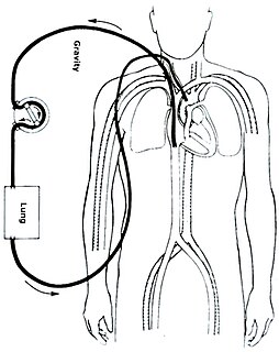
Cardiopulmonary bypass (CPB) is a technique in which a machine temporarily takes over the function of the heart and lungs during surgery, maintaining the circulation of blood and the oxygen content of the patient's body. The CPB pump itself is often referred to as a heart–lung machine or "the pump". Cardiopulmonary bypass pumps are operated by perfusionists. CPB is a form of extracorporeal circulation. Extracorporeal membrane oxygenation is generally used for longer-term treatment.

Pulmonary edema, also known as pulmonary congestion, is excessive liquid accumulation in the tissue and air spaces of the lungs. It leads to impaired gas exchange and may cause hypoxemia and respiratory failure. It is due to either failure of the left ventricle of the heart to remove oxygenated blood adequately from the pulmonary circulation, or an injury to the lung tissue directly or blood vessels of the lung.
Extracorporeal membrane oxygenation (ECMO), also known as extracorporeal life support (ECLS), is an extracorporeal technique of providing prolonged cardiac and respiratory support to persons whose heart and lungs are unable to provide an adequate amount of gas exchange or perfusion to sustain life. The technology for ECMO is largely derived from cardiopulmonary bypass, which provides shorter-term support with arrested native circulation. The device used is a membrane oxygenator, also known as an artificial lung.

Mitral regurgitation(MR), also known as mitral insufficiency or mitral incompetence, is a form of valvular heart disease in which the mitral valve is insufficient and does not close properly when the heart pumps out blood. It is the abnormal leaking of blood backwards – regurgitation from the left ventricle, through the mitral valve, into the left atrium, when the left ventricle contracts. Mitral regurgitation is the most common form of valvular heart disease.

Cardiac catheterization is the insertion of a catheter into a chamber or vessel of the heart. This is done both for diagnostic and interventional purposes.

Cardiogenic shock (CS) is a medical emergency resulting from inadequate blood flow due to the dysfunction of the ventricles of the heart. Signs of inadequate blood flow include low urine production, cool arms and legs, and altered level of consciousness. People may also have a severely low blood pressure and heart rate.

Pulmonary artery catheterization (PAC), or right heart catheterization, is the insertion of a catheter into a pulmonary artery. Its purpose is diagnostic; it is used to detect heart failure or sepsis, monitor therapy, and evaluate the effects of drugs. The pulmonary artery catheter allows direct, simultaneous measurement of pressures in the right atrium, right ventricle, pulmonary artery, and the filling pressure of the left atrium. The pulmonary artery catheter is frequently referred to as a Swan-Ganz catheter, in honor of its inventors Jeremy Swan and William Ganz, from Cedars-Sinai Medical Center.

A ventricular assist device (VAD) is an electromechanical device for assisting cardiac circulation, which is used either to partially or to completely replace the function of a failing heart. The function of a VAD differs from that of an artificial cardiac pacemaker in that a VAD pumps blood, whereas a pacemaker delivers electrical impulses to the heart muscle. Some VADs are for short-term use, typically for patients recovering from myocardial infarction and for patients recovering from cardiac surgery; some are for long-term use, typically for patients suffering from advanced heart failure.
The intra-aortic balloon pump(IABP) is a hail mary mechanical device that increases myocardial oxygen perfusion and indirectly increases cardiac output through afterload reduction. It consists of a cylindrical polyurethane balloon that sits in the aorta, approximately 2 centimeters (0.79 in) from the left subclavian artery. The balloon inflates and deflates via counter pulsation, meaning it actively deflates in systole and inflates in diastole. Systolic deflation decreases afterload through a vacuum effect and indirectly increases forward flow from the heart. Diastolic inflation increases blood flow to the coronary arteries via retrograde flow. These actions combine to decrease myocardial oxygen demand and increase myocardial oxygen supply.

Takotsubo cardiomyopathy or Takotsubo syndrome (TTS), also known as stress cardiomyopathy, is a type of non-ischemic cardiomyopathy in which there is a sudden temporary weakening of the muscular portion of the heart. It usually appears after a significant stressor, either physical or emotional; when caused by the latter, the condition is sometimes called broken heart syndrome. Examples of physical stressors that can cause TTS are sepsis, shock, and pheochromocytoma, and emotional stressors include bereavement, divorce, or the loss of a job. Reviews suggest that of patients diagnosed with the condition, about 70–80% recently experienced a major stressor, including 41–50% with a physical stressor and 26–30% with an emotional stressor. TTS can also appear in patients who have not experienced major stressors.

Myocardial rupture is a laceration of the ventricles or atria of the heart, of the interatrial or interventricular septum, or of the papillary muscles. It is most commonly seen as a serious sequela of an acute myocardial infarction.
The following outline is provided as an overview of and topical guide to cardiology, the branch of medicine dealing with disorders of the human heart. The field includes medical diagnosis and treatment of congenital heart defects, coronary artery disease, heart failure, valvular heart disease and electrophysiology. Physicians who specialize in cardiology are called cardiologists.

Acute decompensated heart failure (ADHF) is a sudden worsening of the signs and symptoms of heart failure, which typically includes difficulty breathing (dyspnea), leg or feet swelling, and fatigue. ADHF is a common and potentially serious cause of acute respiratory distress. The condition is caused by severe congestion of multiple organs by fluid that is inadequately circulated by the failing heart. An attack of decompensation can be caused by underlying medical illness, such as myocardial infarction, an abnormal heart rhythm, infection, or thyroid disease.

Cardiac resynchronisation therapy is the insertion of electrodes in the left and right ventricles of the heart, as well as on occasion the right atrium, to treat heart failure by coordinating the function of the left and right ventricles via a pacemaker, a small device inserted into the interior chest wall.
Thoratec Corporation is a United States-based company that develops, manufactures, and markets proprietary medical devices used for mechanical circulatory support for the treatment of heart-failure patients worldwide. It is a global leader in mechanical circulatory support devices, particularly in ventricular assist devices (VADs).
Myocardial infarction complications may occur immediately following a heart attack, or may need time to develop. After an infarction, an obvious complication is a second infarction, which may occur in the domain of another atherosclerotic coronary artery, or in the same zone if there are any live cells left in the infarct.
Impella is a family of medical devices used for temporary ventricular support in patients with depressed heart function. Some versions of the device can provide left heart support during other forms of mechanical circulatory support including ECMO and Centrimag.

Acute cardiac unloading is any maneuver, therapy, or intervention that decreases the power expenditure of the ventricle and limits the hemodynamic forces that lead to ventricular remodeling after insult or injury to the heart. This technique is being investigated as a therapeutic to aid after damage has occurred to the heart, such as after a heart attack. The theory behind this approach is that by simultaneously limiting the oxygen demand and maximizing oxygen delivery to the heart after damage has occurred, the heart is more fully able to recover. This is primarily achieved by using temporary minimally invasive mechanical circulatory support to supplant the pumping of blood by the heart. Using mechanical support decreases the workload of the heart, or unloads it.
References
- 1 2 3 E.E. Kunst; J.A. van Alste; T. Arts; H.B.K. Boom. ( November, 1994). Integrated Unit for Programmable Control of the 21F Hemopump and registration of Physiological Signs. Medical & Biological Engineering & Computing.
- 1 2 Robert T. Baldwin, MD; Branislav Radovanevic, MD; J. Michael Duncan, MD; Richard K. Wampler, MD; O.H. Frazier, MD (1992). Management of Patients Supported on the Hemopump® Cardiac Assist System. Texas Heart Institute Journal 1992;19:81-6.
- 1 2 He, P. P., Bai, J. J., & Xia, D. D. (2005). Optimum control of the Hemopump as a left-ventricular assist device. Medical & Biological Engineering & Computing, 43(1), 136-141.
- 1 2 3 Texas Heart Institute. (April, 2006). Hemopump.
- ↑ Wampler, Richard; Frazier, O. H. (2018-05-10). "The Hemopump, The First Intravascular Ventricular Assist Device". ASAIO Journal. 65 (3): 297–300. doi: 10.1097/MAT.0000000000000802 . ISSN 1058-2916. PMID 29734260.
- ↑ Urban Lönn, MD; John Wulff, MD; Karl-Yngve Keck, MSEE; Bengt Wranne, MD, PhD; Per Ask, MSEE, PhD; Bengt Peterzén, MD; Henrik Casimir-Ahn, MD, PhD. (January, 1997). Flow Characteristics of the Hemopump: An Experimental In Vitro Study. The Annals of Thoracic Surgery.