
Mastectomy is the medical term for the surgical removal of one or both breasts, partially or completely. A mastectomy is usually carried out to treat breast cancer. In some cases, women believed to be at high risk of breast cancer have the operation as a preventive measure. Alternatively, some women can choose to have a wide local excision, also known as a lumpectomy, an operation in which a small volume of breast tissue containing the tumor and a surrounding margin of healthy tissue is removed to conserve the breast. Both mastectomy and lumpectomy are referred to as "local therapies" for breast cancer, targeting the area of the tumor, as opposed to systemic therapies, such as chemotherapy, hormonal therapy, or immunotherapy.

Plastic surgery is a surgical specialty involving the restoration, reconstruction or alteration of the human body. It can be divided into two main categories: reconstructive surgery and cosmetic surgery. Reconstructive surgery includes craniofacial surgery, hand surgery, microsurgery, and the treatment of burns. While reconstructive surgery aims to reconstruct a part of the body or improve its functioning, cosmetic surgery aims at improving the appearance of it.

A biopsy is a medical test commonly performed by a surgeon, interventional radiologist, or an interventional cardiologist. The process involves extraction of sample cells or tissues for examination to determine the presence or extent of a disease. The tissue is then fixed, dehydrated, embedded, sectioned, stained and mounted before it is generally examined under a microscope by a pathologist; it may also be analyzed chemically. When an entire lump or suspicious area is removed, the procedure is called an excisional biopsy. An incisional biopsy or core biopsy samples a portion of the abnormal tissue without attempting to remove the entire lesion or tumor. When a sample of tissue or fluid is removed with a needle in such a way that cells are removed without preserving the histological architecture of the tissue cells, the procedure is called a needle aspiration biopsy. Biopsies are most commonly performed for insight into possible cancerous or inflammatory conditions.

Eye surgery, also known as ophthalmic or ocular surgery, is surgery performed on the eye or its adnexa, by an ophthalmologist. Eye surgery is part of ophthalmology. The eye is a fragile organ, and requires due care before, during, and after a surgical procedure to minimize or prevent further damage. An eye surgeon is responsible for selecting the appropriate surgical procedure for the patient, and for taking the necessary safety precautions. Mentions of eye surgery can be found in several ancient texts dating back as early as 1800 BC, with cataract treatment starting in the fifth century BC. It continues to be a widely practiced class of surgery, with various techniques having been developed for treating eye problems.

Phacoemulsification is a cataract surgery method in which the internal lens of the eye which has developed a cataract is emulsified with the tip of an ultrasonic handpiece and aspirated from the eye. Aspirated fluids are replaced with irrigation of balanced salt solution to maintain the volume of the anterior chamber during the procedure. This procedure minimises the incision size and reduces the recovery time and risk of surgery induced astigmatism.
Cataract surgery, which is also called lens replacement surgery, is the removal of the natural lens of the human eye that has developed a cataract, an opaque or cloudy area. The eye's natural lens is usually replaced with an artificial intraocular lens (IOL).

A craniotomy is a surgical operation in which a bone flap is temporarily removed from the skull to access the brain. Craniotomies are often critical operations, performed on patients who are suffering from brain lesions, such as tumors, blood clots, removal of foreign bodies such as bullets, or traumatic brain injury (TBI), and can also allow doctors to surgically implant devices, such as deep brain stimulators for the treatment of Parkinson's disease, epilepsy, and cerebellar tremor. The procedure is also used in epilepsy surgery to remove the parts of the brain that are causing epilepsy.
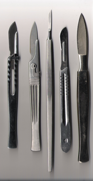
A surgical instrument is a tool or device for performing specific actions or carrying out desired effects during a surgery or operation, such as modifying biological tissue, or to provide access for viewing it. Over time, many different kinds of surgical instruments and tools have been invented. Some surgical instruments are designed for general use in all sorts of surgeries, while others are designed for only certain specialties or specific procedures.

Hair transplantation is a surgical technique that removes hair follicles from one part of the body, called the 'donor site', to a bald or balding part of the body known as the 'recipient site'. The technique is primarily used to treat male pattern baldness. In this minimally invasive procedure, grafts containing hair follicles that are genetically resistant to balding are transplanted to the bald scalp.

A cricothyrotomy is an incision made through the skin and cricothyroid membrane to establish a patent airway during certain life-threatening situations, such as airway obstruction by a foreign body, angioedema, or massive facial trauma. Cricothyrotomy is nearly always performed as a last resort in cases where other means of tracheal intubation are impossible or impractical. Compared with tracheotomy, cricothyrotomy is quicker and easier to perform, does not require manipulation of the cervical spine, and is associated with fewer complications. However, while cricothyrotomy may be life-saving in extreme circumstances, this technique is only intended to be a temporizing measure until a definitive airway can be established.

Mohs surgery, developed in 1938 by a general surgeon, Frederic E. Mohs, is microscopically controlled surgery used to treat both common and rare types of skin cancer. During the surgery, after each removal of tissue and while the patient waits, the tissue is examined for cancer cells. That examination dictates the decision for additional tissue removal. Mohs surgery is the gold standard method for obtaining complete margin control during removal of a skin cancer using frozen section histology. CCPDMA or Mohs surgery allows for the removal of a skin cancer with very narrow surgical margin and a high cure rate.

Veterinary surgery is surgery performed on animals by veterinarians, whereby the procedures fall into three broad categories: orthopaedics, soft tissue surgery, and neurosurgery. Advanced surgical procedures such as joint replacement, fracture repair, stabilization of cranial cruciate ligament deficiency, oncologic (cancer) surgery, herniated disc treatment, complicated gastrointestinal or urogenital procedures, kidney transplant, skin grafts, complicated wound management, and minimally invasive procedures are performed by veterinary surgeons. Most general practice veterinarians perform routine surgeries such as neuters and minor mass excisions; some also perform additional procedures.
Follicular unit transplantation (FUT) is a hair restoration technique, also known as the strip procedure, where a patient's hair is transplanted in naturally occurring groups of 1 to 4 hairs, called follicular units. Follicular units also contain sebaceous (oil) glands, nerves, a small muscle, and occasional fine vellus hairs. In follicular unit transplantation, these small units allow the surgeon to safely transplant thousands of grafts in a single session, which maximizes the cosmetic impact of the procedure.
Follicular unit extraction is one of two primary methods of obtaining hair follicles, naturally occurring groups of one to four hairs, for hair transplantation. The other method is called strip harvesting. In 2018, Mejia published the updated guidelines adopted by the International Society of Hair Restoration Surgery. This name change came about to accurately describe the procedure which involves surgically cutting or incising a full thickness hair follicle skin graft with a circular scalpel, punch or motorized drill and subsequently carefully extracting it from the scalp to be placed in the donor balding scalp. Due to the new developments of incision punches and devices and a variety of different extraction techniques, it was necessary to more accurately define the procedure. Additionally, many places were incorrectly marketing the extraction procedure as a simple plucking of hairs and deceiving the public.
Tubal reversal, also called tubal sterilization reversal, tubal ligation reversal, or microsurgical tubal reanastomosis, is a surgical procedure that can restore fertility to women after a tubal ligation. By rejoining the separated segments of the fallopian tube, tubal reversal can give women the chance to become pregnant again. In some cases, however, the separated segments cannot actually be reattached to each other. In some cases the remaining segment of tube needs to be re-implanted into the uterus. In other cases, when the end of the tube has been removed, a procedure called a neofimbrioplasty must be performed to recreate a functional end of the tube which can then act like the missing fimbria and retrieve the egg that has been released during ovulation.

Penetrating trauma is an open wound injury that occurs when an object pierces the skin and enters a tissue of the body, creating a deep but relatively narrow entry wound. In contrast, a blunt or non-penetrating trauma may have some deep damage, but the overlying skin is not necessarily broken and the wound is still closed to the outside environment. The penetrating object may remain in the tissues, come back out the path it entered, or pass through the full thickness of the tissues and exit from another area.

Skin biopsy is a biopsy technique in which a skin lesion is removed to be sent to a pathologist to render a microscopic diagnosis. It is usually done under local anesthetic in a physician's office, and results are often available in 4 to 10 days. It is commonly performed by dermatologists. Skin biopsies are also done by family physicians, internists, surgeons, and other specialties. However, performed incorrectly, and without appropriate clinical information, a pathologist's interpretation of a skin biopsy can be severely limited, and therefore doctors and patients may forgo traditional biopsy techniques and instead choose Mohs surgery. There are four main types of skin biopsies: shave biopsy, punch biopsy, excisional biopsy, and incisional biopsy. The choice of the different skin biopsies is dependent on the suspected diagnosis of the skin lesion. Like most biopsies, patient consent and anesthesia are prerequisites.
Minimally invasive spine surgery, also known as MISS, has no specific meaning or definition. It implies a lack of severe surgical invasion. The older style of open-spine surgery for a relatively small disc problem used to require a 5-6 inch incision and a month in the hospital. MISS techniques utilize more modern technology, advanced imaging techniques and special medical equipment to reduce tissue trauma, bleeding, radiation exposure, infection risk, and decreased hospital stays by minimizing the size of the incision. Modern endoscopic procedures can be done through a 2 to 5 mm skin opening. By contrast, procedures done with a microscope require skin openings of approximately one inch, or more.

Ancient Roman surgical practices developed from Greek techniques. Roman surgeons and doctors usually learned through apprenticeships or studying. Ancient Roman doctors such as Galen and Celsus described Roman surgical techniques in their medical literature, such as De Medicina. These methods encompassed modern oral surgery, cosmetic surgery, sutures, ligatures, amputations, tonsillectomies, mastectomies, cataract surgeries, lithotomies, hernia repair, gynecology, neurosurgery, and others. Surgery was a rare practice, as it was dangerous and often had fatal results. To perform these procedures, they used tools such as specula, catheters, enemas, bone levers, osteotomes, phlebotomes, probes, curettes, bone drills, bone forceps, cupping vessels, knives, scalpels, scissors, and spathas.
