
The skull is a bone structure that forms the head in vertebrates. It supports the structures of the face and provides a protective cavity for the brain. The skull is composed of two parts: the cranium and the mandible. In humans, these two parts are the neurocranium and the viscerocranium that includes the mandible as its largest bone. The skull forms the anterior-most portion of the skeleton and is a product of cephalisation—housing the brain, and several sensory structures such as the eyes, ears, nose, and mouth. In humans these sensory structures are part of the facial skeleton.

Macrocephaly is a condition in which circumference of the human head is abnormally large. It may be pathological or harmless, and can be a familial genetic characteristic. People diagnosed with macrocephaly will receive further medical tests to determine whether the syndrome is accompanied by particular disorders. Those with benign or familial macrocephaly are considered to have megalencephaly.

Shanidar Cave is an archaeological site located on Bradost Mountain, within the Zagros Mountains, in the Erbil Governorate of Kurdistan Region in northern Iraq. It is known for the discovery of Neanderthal remains at the site most notably Shanidar 1, who survived several injuries during his life, possibly due to care from others in his group, and Shanidar 4, the famed 'flower burial'. Until this discovery, Cro-Magnons, the earliest known H. sapiens in Europe, were the only individuals known for purposeful, ritualistic burials.
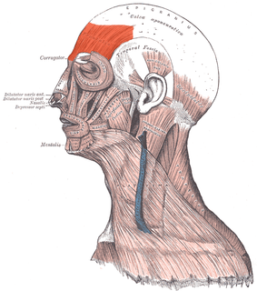
The frontalis muscle is a muscle which covers parts of the forehead of the skull. Some sources consider the frontalis muscle to be a distinct muscle. However, Terminologia Anatomica currently classifies it as part of the occipitofrontalis muscle along with the occipitalis muscle.

The occipitalis muscle is a muscle which covers parts of the skull. Some sources consider the occipital muscle to be a distinct muscle. However, Terminologia Anatomica currently classifies it as part of the occipitofrontalis muscle along with the frontalis muscle.
Craniomandibular osteopathy, also known as lion's jaw, is a developmental disease in dogs causing extensive bony changes in the mandible and skull. In this disease, a cyclical resorption of normal bone and replacement by immature bone occurs along the inner and outer surfaces of the affected bones. It usually occurs between the ages of 3 and 8 months. Breeds most commonly affected include the West Highland White Terrier, Scottish Terrier, Cairn Terrier, and Boston Terrier. It is rare in large-breed dogs, but it has been reported. Symptoms include firm swelling of the jaw, drooling, pain, and difficulty eating.
Infantile cortical hyperostosis is a self-limited inflammatory disorder of infants that causes bone changes, soft tissue swelling and irritability. The disease may be present at birth or occur shortly thereafter. The cause is unknown. Both familial and sporadic forms occur. It is also known as Caffey disease or Caffey's disease.

A frontal eminence is either of two rounded elevations on the frontal bone of the skull. They lie about 3 cm above the supraorbital margin on each side of the frontal suture. They are the site of ossification of the frontal bone during embryological development, although may not be the first site.

Hyperostosis is an excessive growth of bone. It may lead to exostosis. It occurs in many musculoskeletal disorders.
Porotic hyperostosis, is a pathological condition that affects bones of the cranial vault, and is characterized by localized areas of spongy or porous bone tissue. The diploë, or spongy tissue within the bones of the cranium, swells and the tissue of the outer surface becomes thinner and more porous in appearance.
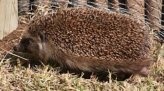
The Southern African hedgehog is a species of mammal in the family Erinaceidae. It is found in Angola, Botswana, Lesotho, Namibia, South Africa, Tanzania and Zimbabwe.
Crista is an internal compartment formed by the inner membrane of a mitochondrion.

Morgagni-Stewart-Morel syndrome is a condition with a wide range of associated endocrine problems including: diabetes mellitus, diabetes insipidus, and hyperparathyroidism. Other signs and symptoms include headaches, vertigo, hirsutism, menstrual disorder, galactorrhoea, obesity, depression, and seizures. Thickening of the inner table of the frontal part of the skull a usually benign condition known as hyperostosis frontalis interna. The syndrome was first described in 1765. It is named after the Italian anatomist and pathologist Giovanni Battista Morgagni, the British neurologist Roy Mackenzie Stewart, and the Swiss psychiatrist Ferdinand Morel.

Diffuse idiopathic skeletal hyperostosis (DISH) is a condition characterized by abnormal calcification/bone formation (hyperostosis) of the soft tissues surrounding the joints of the spine, and also of the peripheral or appendicular skeleton. In the spine, there is bone formation along the anterior longitudinal ligament and sometimes the posterior longitudinal ligament, which may lead to partial or complete fusion of adjacent vertebrae. The facet and sacroiliac joints tend to be uninvolved. The thoracic spine is the most common level involved. In the peripheral skeleton, DISH manifests as a calcific enthesopathy, with pathologic bone formation at sites where ligaments and tendons attach to bone.

Yunnanolepididae is an extinct family of primitive Antiarch placoderms characterized by having short, broad skull roofs, and by having a feature on the visceral side of the posterior medial dorsal plate, the crista transversalis interna posterior, which is diagnostic of antiarchs, turning forward, and lying in front of the posterior ventral process and pit.

Khunnuchelys was a genus of trionychine turtle from the Late Cretaceous of Asia. Three species are known, K. erinhotensis, the type species, K. kizylkumensis, and K. lophorhothon. K. erinhotensis is known from the Iren Dabasu Formation in China from the late Turonian until the middle Campanian. K. kizylkumensis is known from the late Turonian Bissekty Formation of Uzbekistan. The third species, described in 2013 by Danilov et al., is known from the early to middle Campanian aged Bostobe Formation of Kazakhstan.
Van Buchem disease, or hyperostosis corticalis generalisata, is an autosomal recessive skeletal disease which is characterised by uninhibited bone growth, especially in the mandible, skull and ribs.

