The skull is a bone protective cavity for the brain. The skull is composed of four types of bone i.e., cranial bones, facial bones, ear ossicles and hyoid bone, however two parts are more prominent: the cranium and the mandible. In humans, these two parts are the neurocranium (braincase) and the viscerocranium that includes the mandible as its largest bone. The skull forms the anterior-most portion of the skeleton and is a product of cephalisation—housing the brain, and several sensory structures such as the eyes, ears, nose, and mouth. In humans, these sensory structures are part of the facial skeleton.

The jaws are a pair of opposable articulated structure at the entrance of the mouth, typically used for grasping and manipulating food. The term jaws is also broadly applied to the whole of the structures constituting the vault of the mouth and serving to open and close it and is part of the body plan of humans and most animals.

The sphenoid bone is an unpaired bone of the neurocranium. It is situated in the middle of the skull towards the front, in front of the basilar part of the occipital bone. The sphenoid bone is one of the seven bones that articulate to form the orbit. Its shape somewhat resembles that of a butterfly or bat with its wings extended.

The occipital bone is a cranial dermal bone and the main bone of the occiput. It is trapezoidal in shape and curved on itself like a shallow dish. The occipital bone overlies the occipital lobes of the cerebrum. At the base of the skull in the occipital bone, there is a large oval opening called the foramen magnum, which allows the passage of the spinal cord.

The frontal bone or sincipital bone is a bone in the human skull. The bone consists of two portions. These are the vertically oriented squamous part, and the horizontally oriented orbital part, making up the bony part of the forehead, part of the bony orbital cavity holding the eye, and part of the bony part of the nose respectively. The name comes from the Latin word frons.
The parietal bones are two bones in the skull which, when joined at a fibrous joint, form the sides and roof of the cranium. In humans, each bone is roughly quadrilateral in form, and has two surfaces, four borders, and four angles. It is named from the Latin paries (-ietis), wall.

The temporal bones are situated at the sides and base of the skull, and lateral to the temporal lobes of the cerebral cortex.

The vomer is one of the unpaired facial bones of the skull. It is located in the midsagittal line, and articulates with the sphenoid, the ethmoid, the left and right palatine bones, and the left and right maxillary bones. The vomer forms the inferior part of the nasal septum in humans, with the superior part formed by the perpendicular plate of the ethmoid bone. The name is derived from the Latin word for a ploughshare and the shape of the bone.
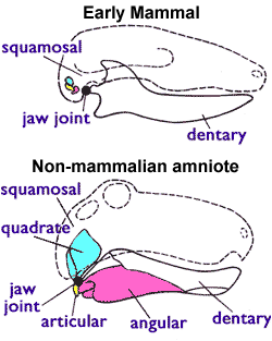
The articular bone is part of the lower jaw of most vertebrates, including most jawed fish, amphibians, birds and various kinds of reptiles, as well as ancestral mammals.

The mastoid part of the temporal bone is the posterior (back) part of the temporal bone, one of the bones of the skull. Its rough surface gives attachment to various muscles and it has openings for blood vessels. From its borders, the mastoid part articulates with two other bones.

The petrous part of the temporal bone is pyramid-shaped and is wedged in at the base of the skull between the sphenoid and occipital bones. Directed medially, forward, and a little upward, it presents a base, an apex, three surfaces, and three angles, and houses in its interior, the components of the inner ear. The petrous portion is among the most basal elements of the skull and forms part of the endocranium. Petrous comes from the Latin word petrosus, meaning "stone-like, hard". It is one of the densest bones in the body. In other mammals, it is a separate bone, the petrosal bone.

The calvaria is the top part of the skull. It is the superior part of the neurocranium and covers the cranial cavity containing the brain. It forms the main component of the skull roof.

In human anatomy, the neurocranium, also known as the braincase, brainpan, or brain-pan, is the upper and back part of the skull, which forms a protective case around the brain. In the human skull, the neurocranium includes the calvaria or skullcap. The remainder of the skull is the facial skeleton.

The premaxilla is one of a pair of small cranial bones at the very tip of the upper jaw of many animals, usually, but not always, bearing teeth. In humans, they are fused with the maxilla. The "premaxilla" of therian mammals has been usually termed as the incisive bone. Other terms used for this structure include premaxillary bone or os premaxillare, intermaxillary bone or os intermaxillare, and Goethe's bone.

Scylacops is an extinct genus of Gorgonopsia. It was first named by Broom in 1913, and contains two species, S. bigendens, and S. capensis. Its fossils have been found in South Africa and Zambia. It is believed to be closely related to the Gorgonopsian Sauroctonus progressus. Scylacops was a moderately sized Gorgonopsid.

The endocranium in comparative anatomy is a part of the skull base in vertebrates and it represents the basal, inner part of the cranium. The term is also applied to the outer layer of the dura mater in human anatomy.

The vertebral column, also known as the backbone or spine, is the core part of the axial skeleton in vertebrate animals. The vertebral column is the defining characteristic of vertebrate endoskeleton in which the notochord found in all chordates has been replaced by a segmented series of mineralized irregular bones called vertebrae, separated by fibrocartilaginous intervertebral discs. The dorsal portion of the vertebral column houses the spinal canal, a cavity formed by alignment of the neural arches that encloses and protects the spinal cord.

Each vertebra is an irregular bone with a complex structure composed of bone and some hyaline cartilage, that make up the vertebral column or spine, of vertebrates. The proportions of the vertebrae differ according to their spinal segment and the particular species.

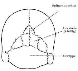
Postparietals are cranial bones present in fish and many tetrapods. Although initially a pair of bones, many lineages possess postparietals which were fused into a single bone. The postparietals were dermal bones situated along the midline of the skull, behind the parietal bones. They formed part of the rear edge of the skull roof, and the lateral edge of each postparietal often contacts the tabular and supratemporal bones. In fish, the postparietals are elongated, typically the largest components of the skull roof. Tetrapods possessed shorter postparietals, which were reduced further and shifted towards the braincase in amniotes. At several points in synapsid evolution, the postparietals fused to each other and the tabulars during embryological development. This fusion produces the interparietal bone, which is inherited by mammals. Postparietals are common in extinct amphibians and early reptiles. However, most living amphibians and living reptiles lack postparietal bones, with a few exceptions.