Intraoral Scanners are devices used in dentistry to capture digital images of the inside of the mouth. These images are an alternative to traditional dental impressions.

Intraoral Scanners are devices used in dentistry to capture digital images of the inside of the mouth. These images are an alternative to traditional dental impressions.
Intraoral Scanners are devices used in dentistry which create digital scans of the teeth and soft tissue anatomy. [1] These devices replace the use of dental putty impressions by using a light source and image sensors to record the tissues inside the mouth accurately and create a virtual alternative to traditional impression plaster models [1]
Dental Impressions are the first step for creating a dental prosthesis. The most common material used for traditional impressions is polyvinyl siloxane, however this material has a poor smell and odour which makes it not favourable for patient or dentist alike. [2] Intraoral scanners have been introduced into dentistry to make the impression process less uncomfortable to the patient. [2]
Intraoral scanners are placed into the mouth and emit a laser or light source which hits the teeth and surrounding tissues, this light is then captured by image sensors and using point clouds, a 3D digital model is made. [1]
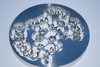
Intraoral Scanners are of high use in CAD/CAM dental use. This is where a computer system can design and mill dental prosthetic framework, such as a crown or bridge, from a digital model. [3] [4]
As these scanners record images of the teeth, they can be used to identify the correct shade for a dental crown. [5]
These devices improve time-management as they show the image in real time. [1] [3] They are also quicker than plaster impressions and more comfortable to the dental patient. [1] [2]
Intraoral scanners have shown to be beneficial to patients suffering from a large gap-reflex which prevents traditional plaster impressions from being taken. [3]
Due to the ‘impressions’ being digital, it means there is no risk of them breaking in transit in comparison with traditional models where they frequently break. [3]
The scanners create a 3D digital scan replicating the intraoral cavity. [4] [2]
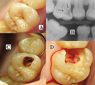
They can detect dental caries, erosion and issues with the periodontium. [4]
Some research has found that digital impressions using intraoral scanners may not be as accurate as traditional plaster impressions [6] [7]

A dentist, also known as a dental surgeon, is a health care professional who specializes in dentistry, the branch of medicine focused on the teeth, gums, and mouth. The dentist's supporting team aids in providing oral health services. The dental team includes dental assistants, dental hygienists, dental technicians, and sometimes dental therapists.
Cosmetic dentistry is generally used to refer to any dental work that improves the appearance of teeth, gums and/or bite. It primarily focuses on improvement in dental aesthetics in color, position, shape, size, alignment and overall smile appearance. Many dentists refer to themselves as "cosmetic dentists" regardless of their specific education, specialty, training, and experience in this field. This has been considered unethical with a predominant objective of marketing to patients. The American Dental Association does not recognize cosmetic dentistry as a formal specialty area of dentistry. However, there are still dentists that promote themselves as cosmetic dentists.

A dental implant is a prosthesis that interfaces with the bone of the jaw or skull to support a dental prosthesis such as a crown, bridge, denture, or facial prosthesis or to act as an orthodontic anchor. The basis for modern dental implants is a biological process called osseointegration, in which materials such as titanium or zirconia form an intimate bond to the bone. The implant fixture is first placed so that it is likely to osseointegrate, then a dental prosthetic is added. A variable amount of healing time is required for osseointegration before either the dental prosthetic is attached to the implant or an abutment is placed which will hold a dental prosthetic or crown.

In dentistry, a crown or a dental cap is a type of dental restoration that completely caps or encircles a tooth or dental implant. A crown may be needed when a large dental cavity threatens the health of a tooth. Some dentists will also finish root canal treatment by covering the exposed tooth with a crown. A crown is typically bonded to the tooth by dental cement. They can be made from various materials, which are usually fabricated using indirect methods. Crowns are used to improve the strength or appearance of teeth and to halt deterioration. While beneficial to dental health, the procedure and materials can be costly.

In dentistry, inlays and onlays are used to fill cavities, and then cemented in place in the tooth. This is an alternative to a direct restoration, made out of composite, amalgam or glass ionomer, that is built up within the mouth.

A dental impression is a negative imprint of hard and soft tissues in the mouth from which a positive reproduction, such as a cast or model, can be formed. It is made by placing an appropriate material in a dental impression tray which is designed to roughly fit over the dental arches. The impression material is liquid or semi-solid when first mixed and placed in the mouth. It then sets to become an elastic solid, which usually takes a few minutes depending upon the material. This leaves an imprint of a person's dentition and surrounding structures of the oral cavity.

An articulator is a mechanical hinged device used in dentistry to which plaster casts of the maxillary (upper) and mandibular (lower) jaw are fixed, reproducing some or all the movements of the mandible in relation to the maxilla. The human maxilla is fixed and the scope of movement of the mandible is dictated by the position and movements of the bilateral temperomandibular joints, which sit in the glenoid fossae in the base of the skull. The temperomandibular joints are not a simple hinge but rotate and translate forward when the mouth is opened.
Orthodontic technology is a specialty of dental technology that is concerned with the design and fabrication of dental appliances for the treatment of malocclusions, which may be a result of tooth irregularity, disproportionate jaw relationships, or both.

CAD/CAM dentistry is a field of dentistry and prosthodontics using CAD/CAM to improve the design and creation of dental restorations, especially dental prostheses, including crowns, crown lays, veneers, inlays and onlays, fixed dental prostheses (bridges), dental implant supported restorations, dentures, and orthodontic appliances. CAD/CAM technology allows the delivery of a well-fitting, aesthetic, and a durable prostheses for the patient. CAD/CAM complements earlier technologies used for these purposes by any combination of increasing the speed of design and creation; increasing the convenience or simplicity of the design, creation, and insertion processes; and making possible restorations and appliances that otherwise would have been infeasible. Other goals include reducing unit cost and making affordable restorations and appliances that otherwise would have been prohibitively expensive. However, to date, chairside CAD/CAM often involves extra time on the part of the dentist, and the fee is often at least two times higher than for conventional restorative treatments using lab services.

Dental radiographs, commonly known as X-rays, are radiographs used to diagnose hidden dental structures, malignant or benign masses, bone loss, and cavities.

Crown lengthening is a surgical procedure performed by a dentist, or more frequently a periodontist, where more tooth is exposed by removing some of the gingival margin (gum) and supporting bone. Crown lengthening can also be achieved orthodontically by extruding the tooth.
In dentistry, overeruption is the physiological movement of a tooth lacking an opposing partner in the dental occlusion. Because of the lack of opposing force and the natural eruptive potential of the tooth there is a tendency for the tooth to erupt out of the line of the occlusion.

Cone beam computed tomography is a medical imaging technique consisting of X-ray computed tomography where the X-rays are divergent, forming a cone.
Air abrasion is a dental technique that uses compressed air to propel a thin stream of abrasive particles—often aluminum oxide or silica—through a specialized hand-piece to remove tooth tissue and decay before being suctioned away, similar to sand blasting. It can be used in a variety of dental procedures, including removing tooth decay, stains, and old restorations, as well as to prepare teeth for new restorations, sealants, and bonding.

Bite registration is a technique carried out in dental procedures, by taking an impression of the teeth, to capture the way the teeth meet together. This is then used to accurately make restorations which will not change the position the teeth meet in.
Digital dentistry refers to the use of dental technologies or devices that incorporates digital or computer-controlled components to carry out dental procedures rather than using mechanical or electrical tools. The use of digital dentistry can make carrying out dental procedures more efficient than using mechanical tools, both for restorative as diagnostic purposes. Used as a way to facilitate dental treatments and propose new ways to meet rising patient demands.
A complete denture is a removable appliance used when all teeth within a jaw have been lost and need to be prosthetically replaced. In contrast to a partial denture, a complete denture is constructed when there are no more teeth left in an arch; hence, it is an exclusively tissue-supported prosthesis. A complete denture can be opposed by natural dentition, a partial or complete denture, fixed appliances or, sometimes, soft tissues.

Overdenture is any removable dental prosthesis that covers and rests on one or more remaining natural teeth, the roots of natural teeth, and/or dental implants. It is one of the most practical measures used in preventive dentistry. Overdentures can be either tooth supported or implant supported. It is found to help in the preservation of alveolar bone and delay the process of complete edentulism.
Occlusion according to The Glossary of Prosthodontic Terms Ninth Edition is defined as "the static relationship between the incising or masticating surfaces of the maxillary or mandibular teeth or tooth analogues".
Full arch restoration in dentistry refers to the comprehensive reconstruction or rehabilitation of an entire dental arch, which can include all teeth in the upper or lower jaw. This procedure is also known as full mouth reconstruction or full mouth rehabilitation.
This article has not been added to any content categories . Please help out by adding categories to it so that it can be listed with similar articles. (September 2024) |