Related Research Articles

Positron emission tomography (PET) is a functional imaging technique that uses radioactive substances known as radiotracers to visualize and measure changes in metabolic processes, and in other physiological activities including blood flow, regional chemical composition, and absorption. Different tracers are used for various imaging purposes, depending on the target process within the body. For example, 18
F
-FDG is commonly used to detect cancer, NaF18
F
is widely used for detecting bone formation, and oxygen-15 is sometimes used to measure blood flow.

Medical imaging is the technique and process of imaging the interior of a body for clinical analysis and medical intervention, as well as visual representation of the function of some organs or tissues (physiology). Medical imaging seeks to reveal internal structures hidden by the skin and bones, as well as to diagnose and treat disease. Medical imaging also establishes a database of normal anatomy and physiology to make it possible to identify abnormalities. Although imaging of removed organs and tissues can be performed for medical reasons, such procedures are usually considered part of pathology instead of medical imaging.

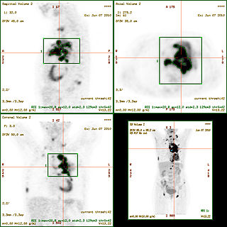
Single-photon emission computed tomography is a nuclear medicine tomographic imaging technique using gamma rays. It is very similar to conventional nuclear medicine planar imaging using a gamma camera, but is able to provide true 3D information. This information is typically presented as cross-sectional slices through the patient, but can be freely reformatted or manipulated as required.

Nuclear medicine is a medical specialty involving the application of radioactive substances in the diagnosis and treatment of disease. Nuclear medicine imaging, in a sense, is "radiology done inside out" or "endoradiology" because it records radiation emitting from within the body rather than radiation that is generated by external sources like X-rays. In addition, nuclear medicine scans differ from radiology, as the emphasis is not on imaging anatomy, but on the function. For such reason, it is called a physiological imaging modality. Single photon emission computed tomography (SPECT) and positron emission tomography (PET) scans are the two most common imaging modalities in nuclear medicine.

A bone scan or bone scintigraphy is a nuclear medicine imaging technique of the bone. It can help diagnose a number of bone conditions, including cancer of the bone or metastasis, location of bone inflammation and fractures, and bone infection (osteomyelitis).

Molecular imaging is a field of medical imaging that focuses on imaging molecules of medical interest within living patients. This is in contrast to conventional methods for obtaining molecular information from preserved tissue samples, such as histology. Molecules of interest may be either ones produced naturally by the body, or synthetic molecules produced in a laboratory and injected into a patient by a doctor. The most common example of molecular imaging used clinically today is to inject a contrast agent into a patient's bloodstream and to use an imaging modality to track its movement in the body. Molecular imaging originated from the field of radiology from a need to better understand fundamental molecular processes inside organisms in a noninvasive manner.

Neuroimaging is the use of quantitative (computational) techniques to study the structure and function of the central nervous system, developed as an objective way of scientifically studying the healthy human brain in a non-invasive manner. Increasingly it is also being used for quantitative studies of brain disease and psychiatric illness. Neuroimaging is a highly multidisciplinary research field and is not a medical specialty.
A gallium scan is a type of nuclear medicine test that uses either a gallium-67 (67Ga) or gallium-68 (68Ga) radiopharmaceutical to obtain images of a specific type of tissue, or disease state of tissue. Gallium salts like gallium citrate and gallium nitrate may be used. The form of salt is not important, since it is the freely dissolved gallium ion Ga3+ which is active. Both 67Ga and 68Ga salts have similar uptake mechanisms. Gallium can also be used in other forms, for example 68Ga-PSMA is used for cancer imaging. The gamma emission of gallium-67 is imaged by a gamma camera, while the positron emission of gallium-68 is imaged by positron emission tomography (PET).
The American College of Radiology (ACR), founded in 1923, is a professional medical society representing nearly 40,000 diagnostic radiologists, radiation oncologists, interventional radiologists, nuclear medicine physicians and medical physicists.
Perfusion is the passage of fluid through the lymphatic system or blood vessels to an organ or a tissue. The practice of perfusion scanning is the process by which this perfusion can be observed, recorded and quantified. The term perfusion scanning encompasses a wide range of medical imaging modalities.
Nuclear medicine physicians, also called nuclear radiologists, are medical specialists that use tracers, usually radiopharmaceuticals, for diagnosis and therapy. Nuclear medicine procedures are the major clinical applications of molecular imaging and molecular therapy. In the United States, nuclear medicine physicians are certified by the American Board of Nuclear Medicine and the American Osteopathic Board of Nuclear Medicine.
The standardized uptake value (SUV) is a nuclear medicine term, used in positron emission tomography (PET) as well as in modern calibrated single photon emission tomography (SPECT) imaging for a semiquantitative analysis. Its use is particularly common in the analysis of [18F]fluorodeoxyglucose ([18F]FDG) images of cancer patients. It can also be used with other PET agents especially when no arterial input function is available for more detailed pharmacokinetic modeling. Otherwise measures like the fractional uptake rate (FUR) or parameters from more advanced pharmacokinetic modeling may be preferable.
Preclinical imaging is the visualization of living animals for research purposes, such as drug development. Imaging modalities have long been crucial to the researcher in observing changes, either at the organ, tissue, cell, or molecular level, in animals responding to physiological or environmental changes. Imaging modalities that are non-invasive and in vivo have become especially important to study animal models longitudinally. Broadly speaking, these imaging systems can be categorized into primarily morphological/anatomical and primarily molecular imaging techniques. Techniques such as high-frequency micro-ultrasound, magnetic resonance imaging (MRI) and computed tomography (CT) are usually used for anatomical imaging, while optical imaging, positron emission tomography (PET), and single photon emission computed tomography (SPECT) are usually used for molecular visualizations.

Imaging phantom, or simply phantom, is a specially designed object that is scanned or imaged in the field of medical imaging to evaluate, analyze, and tune the performance of various imaging devices. A phantom is more readily available and provides more consistent results than the use of a living subject or cadaver, and likewise avoids subjecting a living subject to direct risk. Phantoms were originally employed for use in 2D x-ray based imaging techniques such as radiography or fluoroscopy, though more recently phantoms with desired imaging characteristics have been developed for 3D techniques such as SPECT, MRI, CT, Ultrasound, PET, and other imaging methods or modalities.

Positron emission tomography–magnetic resonance imaging (PET–MRI) is a hybrid imaging technology that incorporates magnetic resonance imaging (MRI) soft tissue morphological imaging and positron emission tomography (PET) functional imaging.
Computational human phantoms are models of the human body used in computerized analysis. Since the 1960s, the radiological science community has developed and applied these models for ionizing radiation dosimetry studies. These models have become increasingly accurate with respect to the internal structure of the human body.

Cardiac imaging refers to non-invasive imaging of the heart using ultrasound, magnetic resonance imaging (MRI), computed tomography (CT), or nuclear medicine (NM) imaging with PET or SPECT. These cardiac techniques are otherwise referred to as echocardiography, Cardiac MRI, Cardiac CT, Cardiac PET and Cardiac SPECT including myocardial perfusion imaging.
Preclinical or small-animal Single Photon Emission Computed Tomography (SPECT) is a radionuclide based molecular imaging modality for small laboratory animals. Although SPECT is a well-established imaging technique that is already for decades in use for clinical application, the limited resolution of clinical SPECT (~10 mm) stimulated the development of dedicated small animal SPECT systems with sub-mm resolution. Unlike in clinics, preclinical SPECT outperforms preclinical coincidence PET in terms of resolution and, at the same time, allows to perform fast dynamic imaging of animals.
Sandip Basu is an Indian physician of Nuclear Medicine and the Head, Nuclear Medicine Academic Program at the Radiation Medicine Centre. He is also the Dean-Academic (Health-Sciences), BARC at Homi Bhabha National Institute and is known for his services and research in Nuclear Medicine, particularly on Positron emission tomography diagnostics and Targeted Radionuclide Therapy in Cancer. The Council of Scientific and Industrial Research, the apex agency of the Government of India for scientific research, awarded him the Shanti Swarup Bhatnagar Prize for Science and Technology, one of the highest Indian science awards for his contributions to Nuclear Medicine in 2012.
Philip F. Cohen is a Canadian clinical director of Nuclear Medicine working out of the Lions Gate Hospital in North Vancouver, British Columbia. As a nuclear medicine physician, he is a pioneer in the usage of 3-D imaging techniques to improve diagnosis of bone disease and injury in collaboration with the Medical Imaging Research Group at University of British Columbia. Furthermore, Cohen has been involved in clinical research trials of new radiopharmaceuticals. To that effect, Cohen was the first recipient of a research grant from the Lions Gate Hospital Foundation, one of several peer-reviewed awards that would follow.
References
- ↑ Quality assurance for SPECT systems. IAEA Health Series No. 6. 2009. International Atomic Energy Agency publications. ISBN 978-92-0-103709-1 p.182
- ↑ MacFarlane, Carolyn Richards; American College of Radiologists (March 2006). "ACR Accreditation of Nuclear Medicine and PET Imaging Departments". Journal of Nuclear Medicine Technology. 34 (1): 18–24. PMID 16517965.
- ↑ "ACR Nuclear Medicine & PET Accreditation".
- ↑ Jennifer Prekeges. Nuclear Medicine Instrumentation. Jones & Bartlett Publishers. 2012. ISBN 1449645372 p.189
- ↑ "Ronald Jaszczak, PhD". SNMMI Annual Meeting. Archived from the original on 2016-10-11. Retrieved 2016-07-02.
- ↑ USPATENT 4499375,Ronald J Jaszczak,"Nuclear imaging phantom",issued 1985-02-12
- ↑ Mattsson S, Hoeschen C. Radiation Protection in Nuclear Medicine. Springer. 2003. ISBN 978-3-642-31166-6. p.82
- ↑ Waterstram-Rich KM, Christian PE. Nuclear Medicine and PET/CT. 7th Ed. Elsevier Health Sciences, 2013. ISBN 0323277047 p.345
- ↑ Bolus NE, Brady AB. Steves' Review of Nuclear Medicine Technology. Society of Nuclear Medicine. 4th Ed. 2011. ISBN 978-0-932004-87-1 p.177
- ↑ Jaszczak, Ronald Jack (7 July 2006). "The early years of single photon emission computed tomography (SPECT): an anthology of selected reminiscences" (PDF). Physics in Medicine and Biology. 51 (13): R99–R115. CiteSeerX 10.1.1.456.9131 . doi:10.1088/0031-9155/51/13/R07. PMID 16790923.
- ↑ "Phantom Testing: Nuclear Medicine". Accreditation Support. American College of Radiology. 4 February 2021. Retrieved 8 March 2022.
- ↑ Bailey DL, Humm JL, et al. Nuclear Medicine Physics: A Handbook for Teachers and Students. 2014. International Atomic Energy Agency publications. ISBN 978-92-0-143810-2. p.563