Method

The test combines a test plasma with kaolin, and after a brief pre-incubation and the addition of calcium chloride, the time to clot (in seconds) is measured. [6] Mixes of patient plasma with normal plasma are recommended for testing. [9]
| Kaolin clotting time | |
|---|---|
| Test of | Blood plasma [1] |
Kaolin clotting time (KCT) is a sensitive test to detect lupus anticoagulants. [2] There is evidence that suggests it is the most sensitive test for detecting lupus anticoagulants. [3] It can also detect factor VIII inhibitors but is sensitive to unfractionated heparin as well. [4]
The KCT on whole blood is known as the "Activated Clotting Time" (ACT) and is widely used in various instruments during surgery such as cardiac bypass to monitor heparin. [5]
KCT was first described by Dr. Joel Margolis in 1958. [6] Later on, it was found to be very sensitive to lupus anticoagulants but was only reliable when test plasmas were mixed with normal plasma in various proportions. [3] It became the preferred method for lupus anticoagulant testing after Dr. Wilhelm Lubbe showed it to be a good marker for recurrent fetal loss. [7]
KCT is similar to the activated partial thromboplastin time test, except it does not use exogenous phospholipid. [2] Thus, a confirmatory test that uses excess phospholipid is needed to validate the presence of lupus anticoagulants. [2] Otherwise, diluting the test plasma in normal plasma before testing provides characteristic mixing patterns. [8]
Kaolin is the surface activator, and the test also requires small amounts of cell fragments and plasma lipids to provide the phospholipid surface required for coagulation. [2] [4] Therefore, the sample quality is important for the validity of the screening test. [2]
The test combines a test plasma with kaolin, and after a brief pre-incubation and the addition of calcium chloride, the time to clot (in seconds) is measured. [6] Mixes of patient plasma with normal plasma are recommended for testing. [9]
The KCT test/control ratio of greater than or equal to 1.2 indicates that a defect is present. [4] If the test/control ratio is between 1.1 and 1.2, the test is equivocal. [4]
A good way of expressing the result using mixes is to calculate the Rosner index. [10] If A is the KCT of normal plasma, B is that of the 1:1 mix and C is that of the patient plasma, then the Rosner index is 100x(B-A)/C. Values above 15 indicate a positive result but in most cases labs set their own cutoff values. [9]
If the KCT is less than 60 seconds, this suggests that the test plasma is contaminated with platelet fragments; therefore, the test is not valid. [4]

Coagulation, also known as clotting, is the process by which blood changes from a liquid to a gel, forming a blood clot. It potentially results in hemostasis, the cessation of blood loss from a damaged vessel, followed by repair. The mechanism of coagulation involves activation, adhesion and aggregation of platelets, as well as deposition and maturation of fibrin.

Disseminated intravascular coagulation (DIC) is a condition in which blood clots form throughout the body, blocking small blood vessels. Symptoms may include chest pain, shortness of breath, leg pain, problems speaking, or problems moving parts of the body. As clotting factors and platelets are used up, bleeding may occur. This may include blood in the urine, blood in the stool, or bleeding into the skin. Complications may include organ failure.
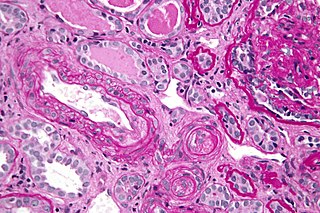
Antiphospholipid syndrome, or antiphospholipid antibody syndrome, is an autoimmune, hypercoagulable state caused by antiphospholipid antibodies. APS provokes blood clots (thrombosis) in both arteries and veins as well as pregnancy-related complications such as miscarriage, stillbirth, preterm delivery, and severe preeclampsia. Although the exact etiology of APS is still not clear, genetics is believed to play a key role in the development of the disease. The diagnostic criteria require one clinical event and two positive blood test results spaced at least three months apart that detect lupus anticoagulant, anti-apolipoprotein antibodies, or anti-cardiolipin antibodies.

Fibrinogen is a glycoprotein complex, produced in the liver, that circulates in the blood of all vertebrates. During tissue and vascular injury, it is converted enzymatically by thrombin to fibrin and then to a fibrin-based blood clot. Fibrin clots function primarily to occlude blood vessels to stop bleeding. Fibrin also binds and reduces the activity of thrombin. This activity, sometimes referred to as antithrombin I, limits clotting. Fibrin also mediates blood platelet and endothelial cell spreading, tissue fibroblast proliferation, capillary tube formation, and angiogenesis and thereby promotes revascularization and wound healing.

Haemophilia C (also known as plasma thromboplastin antecedent deficiency or Rosenthal syndrome) is a mild form of haemophilia affecting both sexes, due to factor XI deficiency. It predominantly occurs in Ashkenazi Jews. It is the fourth most common coagulation disorder after von Willebrand's disease and haemophilia A and B. In the United States, it is thought to affect 1 in 100,000 of the adult population, making it 10% as common as haemophilia A.
Low-molecular-weight heparin (LMWH) is a class of anticoagulant medications. They are used in the prevention of blood clots and treatment of venous thromboembolism and in the treatment of myocardial infarction.

The prothrombin time (PT) – along with its derived measures of prothrombin ratio (PR) and international normalized ratio (INR) – is an assay for evaluating the extrinsic pathway and common pathway of coagulation. This blood test is also called protime INR and PT/INR. They are used to determine the clotting tendency of blood, in such things as the measure of warfarin dosage, liver damage, and vitamin K status. PT measures the following coagulation factors: I (fibrinogen), II (prothrombin), V (proaccelerin), VII (proconvertin), and X.

The partial thromboplastin time (PTT), also known as the activated partial thromboplastin time, is a blood test that characterizes coagulation of the blood. A historical name for this measure is the kaolin-cephalin clotting time (KCCT), reflecting kaolin and cephalin as materials historically used in the test. Apart from detecting abnormalities in blood clotting, partial thromboplastin time is also used to monitor the treatment effect of heparin, a widely prescribed drug that reduces blood's tendency to clot.
Mixing studies are tests performed on blood plasma of patients or test subjects to distinguish factor deficiencies from factor inhibitors, such as lupus anticoagulant, or specific factor inhibitors, such as antibodies directed against factor VIII. The basic purpose of these tests is to determine the cause of prolongation of Prothrombin Time (PT), Partial Thromboplastin Time, or sometimes of thrombin time (TT). Mixing studies take advantage of the fact that factor levels that are 50 percent of normal should give a normal Prothrombin time (PT) or Partial thromboplastin time (PTT) result. Factor deficient plasmas are used in mixing studies. Plasma with known factor deficiencies are commercially available but are very expensive, so they are often prepared in the laboratory and can then be used for mixing experiments.
Lupus anticoagulant is an immunoglobulin that binds to phospholipids and proteins associated with the cell membrane. Its name is a partial misnomer, as it is actually a prothrombotic antibody in vivo. Lupus anticoagulant in living systems causes a decrease in clotting time. The name derives from their properties in vitro, as these antibodies increase coagulation times in laboratory tests such as the activated partial thromboplastin time (aPTT). Investigators speculate that the antibodies interfere with phospholipids used to induce in vitro coagulation. In vivo, the antibodies are thought to interact with platelet membrane phospholipids, increasing adhesion and aggregation of platelets, which accounts for the in vivo prothrombotic characteristics.

Dilute Russell's viper venom time (dRVVT) is a laboratory test often used for detection of lupus anticoagulant (LA).
Tissue factor, also called platelet tissue factor, factor III, or CD142, is a protein encoded by the F3 gene, present in subendothelial tissue and leukocytes. Its role in the clotting process is the initiation of thrombin formation from the zymogen prothrombin. Thromboplastin defines the cascade that leads to the activation of factor X—the tissue factor pathway. In doing so, it has replaced the previously named extrinsic pathway in order to eliminate ambiguity.
Thromboelastography (TEG) is a method of testing the efficiency of blood coagulation. It is a test mainly used in surgery and anesthesiology, although increasingly used in resuscitations in Emergency Departments, intensive care units, and labor and delivery suites. More common tests of blood coagulation include prothrombin time (PT) and partial thromboplastin time (aPTT) which measure coagulation factor function, but TEG also can assess platelet function, clot strength, and fibrinolysis which these other tests cannot.
The thrombin time (TT), also known as the thrombin clotting time (TCT), is a blood test that measures the time it takes for a clot to form in the plasma of a blood sample containing anticoagulant, after an excess of thrombin has been added. It is used to diagnose blood coagulation disorders and to assess the effectiveness of fibrinolytic therapy. This test is repeated with pooled plasma from normal patients. The difference in time between the test and the 'normal' indicates an abnormality in the conversion of fibrinogen to fibrin, an insoluble protein.

Ecarin clotting time (ECT) is a laboratory test used to monitor anticoagulation during treatment with hirudin, an anticoagulant medication which was originally isolated from leech saliva. Ecarin, the primary reagent in this assay, is derived from the venom of the saw-scaled viper, Echis carinatus.
Thromboelastometry (TEM), previously named rotational thromboelastography (ROTEG) or rotational thromboelastometry (ROTEM), is an established viscoelastic method for hemostasis testing in whole blood. It is a modification of traditional thromboelastography (TEG).
Activated clotting time (ACT), also known as activated coagulation time, is a test of coagulation.
Thrombodynamics test is a method for blood coagulation monitoring and anticoagulant control. This test is based on imitation of coagulation processes occurring in vivo, is sensitive both to pro- and anticoagulant changes in the hemostatic balance. Highly sensitive to thrombosis.
Blood clotting tests are the tests used for diagnostics of the hemostasis system. Coagulometer is the medical laboratory analyzer used for testing of the hemostasis system. Modern coagulometers realize different methods of activation and observation of development of blood clots in blood or in blood plasma.
The haemostatic system involves the interaction of proteins in the blood, the blood vessel wall and the flow of blood to control bleeding and blood clotting. Developmental Haemostasis is a term that represents the maturation of the haemostatic system from birth to adulthood. There are differences in the concentration, structure and activity of many proteins involved in blood clotting. These changes play an important role in physiological development and are important in providing appropriate diagnosis and treatment of bleeding and clotting disorders. The age-specific differences in the blood clotting system may contribute to the fact that children are less prone to developing thrombosis compared to adults.