Related Research Articles
An electron microscope is a microscope that uses a beam of accelerated electrons as a source of illumination. As the wavelength of an electron can be up to 100,000 times shorter than that of visible light photons, electron microscopes have a higher resolving power than light microscopes and can reveal the structure of smaller objects. A scanning transmission electron microscope has achieved better than 50 pm resolution in annular dark-field imaging mode and magnifications of up to about 10,000,000× whereas most light microscopes are limited by diffraction to about 200 nm resolution and useful magnifications below 2000×.

Histology, also known as microscopic anatomy or microanatomy, is the branch of biology which studies the microscopic anatomy of biological tissues. Histology is the microscopic counterpart to gross anatomy, which looks at larger structures visible without a microscope. Although one may divide microscopic anatomy into organology, the study of organs, histology, the study of tissues, and cytology, the study of cells, modern usage places these topics under the field of histology. In medicine, histopathology is the branch of histology that includes the microscopic identification and study of diseased tissue. In the field of paleontology, the term paleohistology refers to the histology of fossil organisms.

Microscopy is the technical field of using microscopes to view objects and areas of objects that cannot be seen with the naked eye. There are three well-known branches of microscopy: optical, electron, and scanning probe microscopy, along with the emerging field of X-ray microscopy.

Positron emission tomography (PET) is a functional imaging technique that uses radioactive substances known as radiotracers to visualize and measure changes in metabolic processes, and in other physiological activities including blood flow, regional chemical composition, and absorption. Different tracers are used for various imaging purposes, depending on the target process within the body. For example, 18F-FDG is commonly used to detect cancer, NaF-F18 is widely used for detecting bone formation, and oxygen-15 is sometimes used to measure blood flow.
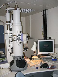
A scanning electron microscope (SEM) is a type of electron microscope that produces images of a sample by scanning the surface with a focused beam of electrons. The electrons interact with atoms in the sample, producing various signals that contain information about the surface topography and composition of the sample. The electron beam is scanned in a raster scan pattern, and the position of the beam is combined with the intensity of the detected signal to produce an image. In the most common SEM mode, secondary electrons emitted by atoms excited by the electron beam are detected using a secondary electron detector. The number of secondary electrons that can be detected, and thus the signal intensity, depends, among other things, on specimen topography. Some SEMs can achieve resolutions better than 1 nanometer.
Phosphatic fossilization has occurred in unusual circumstances to preserve some extremely high-resolution microfossils in which careful preparation can even reveal preserved cellular structures. Such microscopic fossils are only visible under the scanning electron microscope.

Optical coherence tomography (OCT) is an imaging technique that uses low-coherence light to capture micrometer-resolution, two- and three-dimensional images from within optical scattering media. It is used for medical imaging and industrial nondestructive testing (NDT). Optical coherence tomography is based on low-coherence interferometry, typically employing near-infrared light. The use of relatively long wavelength light allows it to penetrate into the scattering medium. Confocal microscopy, another optical technique, typically penetrates less deeply into the sample but with higher resolution.

An X-ray microscope uses electromagnetic radiation in the soft X-ray band to produce magnified images of objects. Since X-rays penetrate most objects, there is no need to specially prepare them for X-ray microscopy observations.

Confocal microscopy, most frequently confocal laser scanning microscopy (CLSM) or laser confocal scanning microscopy (LCSM), is an optical imaging technique for increasing optical resolution and contrast of a micrograph by means of using a spatial pinhole to block out-of-focus light in image formation. Capturing multiple two-dimensional images at different depths in a sample enables the reconstruction of three-dimensional structures within an object. This technique is used extensively in the scientific and industrial communities and typical applications are in life sciences, semiconductor inspection and materials science.

Microsurgery is a general term for surgery requiring an operating microscope. The most obvious developments have been procedures developed to allow anastomosis of successively smaller blood vessels and nerves which have allowed transfer of tissue from one part of the body to another and re-attachment of severed parts. Microsurgical techniques are utilized by several specialties today, such as: general surgery, ophthalmology, orthopedic surgery, gynecological surgery, otolaryngology, neurosurgery, oral and maxillofacial surgery, plastic surgery, podiatric surgery and pediatric surgery.
A microtome is a tool used to cut extremely thin slices of material, known as sections. Important in science, microtomes are used in microscopy, allowing for the preparation of samples for observation under transmitted light or electron radiation.
Winfried Denk built the first two-photon microscope while he was a graduate student in Watt W. Webb's lab at Cornell University, in 1989.
Bruce Howard McCormick (1928–2007) was an American computer scientist, Emeritus Professor at the Department of Computer Science, and founding director of the Brain Networks Lab at Texas A&M University.
Christoph Cremer is a German physicist and emeritus at the Ruprecht-Karls-University Heidelberg, honorary professor at the University of Mainz and was a former group leader at the Institute of Molecular Biology (IMB) at the Johannes Gutenberg University of Mainz, Germany, who has successfully overcome the conventional limit of resolution that applies to light based investigations by a range of different methods.
Serial block-face scanning electron microscopy is a method to generate high resolution three-dimensional images from small samples. The technique was developed for brain tissue, but it is widely applicable for any biological samples. A serial block-face scanning electron microscope consists of an ultramicrotome mounted inside the vacuum chamber of a scanning electron microscope. Samples are prepared by methods similar to that in transmission electron microscopy (TEM), typically by fixing the sample with aldehyde, staining with heavy metals such as osmium and uranium then embedding in an epoxy resin. The surface of the block of resin-embedded sample is imaged by detection of back-scattered electrons. Following imaging the ultramicrotome is used to cut a thin section from the face of the block. After the section is cut, the sample block is raised back to the focal plane and imaged again. This sequence of sample imaging, section cutting and block raising can acquire many thousands of images in perfect alignment in an automated fashion. Practical serial block-face scanning electron microscopy was invented in 2004 by Winfried Denk at the Max-Planck-Institute in Heidelberg and is commercially available from Gatan Inc., Thermo Fisher Scientific (VolumeScope) and ConnectomX.
Endomicroscopy is a technique for obtaining histology-like images from inside the human body in real-time, a process known as ‘optical biopsy’. It generally refers to fluorescence confocal microscopy, although multi-photon microscopy and optical coherence tomography have also been adapted for endoscopic use. Commercially available clinical and pre-clinical endomicroscopes can achieve a resolution on the order of a micrometre, have a field-of-view of several hundred µm, and are compatible with fluorophores which are excitable using 488 nm laser light. The main clinical applications are currently in imaging of the tumour margins of the brain and gastro-intestinal tract, particularly for the diagnosis and characterisation of Barrett’s Esophagus, pancreatic cysts and colorectal lesions. A number of pre-clinical and transnational applications have been developed for endomicroscopy as it enables researchers to perform live animal imaging. Major pre-clinical applications are in gastro-intestinal tract, toumour margin detection, uterine complications, ischaemia, live imaging of cartilage and tendon and organoid imaging.

Todd Huffman is an American technology entrepreneur and prolific photographer. He is a co-founder of the biomedical imaging company, 3Scan.
3Scan, Inc. is an American biotechnology company based in San Francisco, California. It offers automated microscopy services using a coordinated combination of both hardware and software for the 3D analysis of cells, tissues, and organs. The company was founded in 2011 by Todd Huffman, Megan Klimen, Matthew Goodman, and Cody Daniel. The 3Scan technology is based on the Knife Edge Scanning Microscope developed in the late 1990s by Bruce McCormick, founder of the Brain Networks Lab at Texas A&M University.
Three-photon microscopy (3PEF) is a high-resolution fluorescence microscopy based on nonlinear excitation effect. Different from two photon excitation microscopy, it uses three exciting photons. It typically uses 1300nm or longer wavelength laser to excite the fluorescent dyes with three simultaneously absorbed photons, and then the fluorescent dyes emit one photon whose energy is three times the energy of each incident photon. Comparing to two-photon microscopy, three-photon microscopy reduces the fluorescence away from the focal plan by , which is much faster than that of two-photon microscopy. In addition, three-photon microscopy employs near-infrared light with less tissue scattering effect, which causes three photon microscopy to have higher resolution than conventional microscopy.

Gail McConnell is a Scottish physicist who is Professor of Physics and director of the Centre for Biophotonics at the University of Strathclyde. She is interested in optical microscopy and novel imaging techniques, and leads the Mesolens microscope facility where her research investigates linear and non-linear optics.
References
- ↑ Kathy Flores (2007). "Dr. Bruce H. McCormick" Texas A&M University. Retrieved 9 July 2008.
- ↑ B. H. McCormick, Development of the Brain Tissue Scanner, Technical Report, Brain Networks Laboratory, Department of Computer Science, Texas A&M University, College Station, Texas, 2002. [PDF]
- ↑ Knife-edge scanning microscope (KESM): http://research.cs.tamu.edu/bnl/kesm.html