The frontal bone is a bone in the human skull. The bone consists of two portions. These are the vertically oriented squamous part, and the horizontally oriented orbital part, making up the bony part of the forehead, part of the bony orbital cavity holding the eye, and part of the bony part of the nose respectively. The name comes from the Latin word frons.

The lacrimal bone is a small and fragile bone of the facial skeleton; it is roughly the size of the little fingernail. It is situated at the front part of the medial wall of the orbit. It has two surfaces and four borders. Several bony landmarks of the lacrimal bone function in the process of lacrimation or crying. Specifically, the lacrimal bone helps form the nasolacrimal canal necessary for tear translocation. A depression on the anterior inferior portion of the bone, the lacrimal fossa, houses the membranous lacrimal sac. Tears or lacrimal fluid, from the lacrimal glands, collect in this sac during excessive lacrimation. The fluid then flows through the nasolacrimal duct and into the nasopharynx. This drainage results in what is commonly referred to a runny nose during excessive crying or tear production. Injury or fracture of the lacrimal bone can result in posttraumatic obstruction of the lacrimal pathways.

In anatomy, the orbit is the cavity or socket of the skull in which the eye and its appendages are situated. "Orbit" can refer to the bony socket, or it can also be used to imply the contents. In the adult human, the volume of the orbit is 30 millilitres, of which the eye occupies 6.5 ml. The orbital contents comprise the eye, the orbital and retrobulbar fascia, extraocular muscles, cranial nerves II, III, IV, V, and VI, blood vessels, fat, the lacrimal gland with its sac and duct, the eyelids, medial and lateral palpebral ligaments, check ligaments, the suspensory ligament, septum, ciliary ganglion and short ciliary nerves.

The lacrimal glands are paired exocrine glands, one for each eye, found in most terrestrial vertebrates and some marine mammals, that secrete the aqueous layer of the tear film. In humans, they are situated in the upper lateral region of each orbit, in the lacrimal fossa of the orbit formed by the frontal bone. Inflammation of the lacrimal glands is called dacryoadenitis. The lacrimal gland produces tears which are secreted by the lacrimal ducts, and flow over the ocular surface, and then into canals that connect to the lacrimal sac. From that sac, the tears drain through the lacrimal duct into the nose.

The orbital or horizontal part of the frontal bone consists of two thin triangular plates, the orbital plates, which form the vaults of the orbits, and are separated from one another by a median gap, the ethmoidal notch.

The tarsi are two comparatively thick, elongated plates of dense connective tissue, about 10 mm (0.39 in) in length for the upper eyelid and 5 mm for the lower eyelid; one is found in each eyelid, and contributes to its form and support. They are located directly above the lid margins. The tarsus has a lower and upper part making up the palpebrae.

The lacrimal nerve is the smallest branch of the ophthalmic nerve (V1), itself a branch of the trigeminal nerve (CN V). The other branches of the ophthalmic nerve are the frontal nerve and nasociliary nerve.

The greater wing of the sphenoid bone, or alisphenoid, is a bony process of the sphenoid bone; there is one on each side, extending from the side of the body of the sphenoid and curving upward, laterally, and backward.

The ethmoidal labyrinth or lateral mass of the ethmoid bone consists of a number of thin-walled cellular cavities, the ethmoid air cells, arranged in three groups, anterior, middle, and posterior, and interposed between two vertical plates of bone; the lateral plate forms part of the orbit, the medial plate forms part of the nasal cavity. In the disarticulated bone many of these cells are opened into, but when the bones are articulated, they are closed in at every part, except where they open into the nasal cavity.
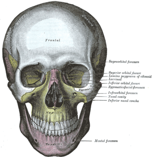
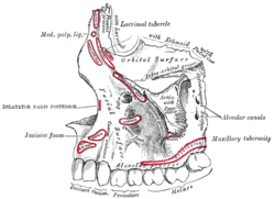
In human anatomy, the infraorbital foramen is an opening in the maxillary bone of the skull located below the infraorbital margin of the orbit. It transmits the infraorbital artery and vein, and the infraorbital nerve, a branch of the maxillary nerve. It is typically 6.10 to 10.9 mm from the infraorbital margin.

The lacrimal artery is an artery of the orbit around the eye. It arises close to the optic foramen, and is a branch of the ophthalmic artery. It accompanies the lacrimal nerve along the upper border of the lateral rectus muscle. It supplies the lacrimal gland, two rectus muscles of the eye, the eyelids, and the conjunctiva.

The posterior lacrimal crest is a vertical bony ridge on the orbital surface of the lacrimal bone. It divides the bone into two parts. It gives origin to the lacrimal part of the orbicularis oculi muscle.
The anterior lacrimal crest is a bony projection on the frontal process of the maxilla. It creates the lateral margin of the lacrimal sac fossa and is continuous with the orbital margin. The medial palpebral ligament is attached to anterior lacrimal crest. It is an important structure to avoid damaging during rhinoplasty.

The zygomatic processes are three processes (protrusions) from other bones of the skull which each articulate with the zygomatic bone. The three processes are:

The frontal process of maxilla is a strong plate, which projects upward, medialward, and backward from the maxilla, forming part of the lateral boundary of the nose.
The medial palpebral arteries are arteries of the head. They are two in number, superior and inferior, that arise from the ophthalmic artery near its terminal branches. The arteries branch from the ophthalmic artery in the superomedial orbit near the trochlea of the superior oblique muscle.
The Orbital Fascia forms the periosteum of the orbit.

The following outline is provided as an overview of and topical guide to human anatomy:
The lacrimal hamulus is a small, hook-like bony projection of the lacrimal bone. It is a continuation of the posterior lacrimal crest. It articulates with the lacrimal tubercle of the maxilla, and completes the upper orifice of the lacrimal canaliculus. It sometimes exists as a separate piece, and is then called the lesser lacrimal bone.

The accessory visual structures are the protecting and supporting structures (adnexa) of the eye, including the eyebrow, eyelids, and lacrimal apparatus. The eyebrows, eyelids, eyelashes, lacrimal gland and drainage apparatus all play a crucial role with regards to globe protection, lubrication, and minimizing the risk of ocular infection. The adnexal structures also help to keep the cornea moist and clean.