Related Research Articles

In vertebrate anatomy, ribs are the long curved bones which form the rib cage, part of the axial skeleton. In most tetrapods, ribs surround the thoracic cavity, enabling the lungs to expand and thus facilitate breathing by expanding the thoracic cavity. They serve to protect the lungs, heart, and other vital organs of the thorax. In some animals, especially snakes, ribs may provide support and protection for the entire body.

The rib cage or thoracic cage is an endoskeletal enclosure in the thorax of most vertebrates that comprises the ribs, vertebral column and sternum, which protect the vital organs of the thoracic cavity, such as the heart, lungs and great vessels and support the shoulder girdle to form the core part of the axial skeleton.

The clavicle, collarbone, or keybone is a slender, S-shaped long bone approximately 6 inches (15 cm) long that serves as a strut between the shoulder blade and the sternum (breastbone). There are two clavicles, one on each side of the body. The clavicle is the only long bone in the body that lies horizontally. Together with the shoulder blade, it makes up the shoulder girdle. It is a palpable bone and, in people who have less fat in this region, the location of the bone is clearly visible. It receives its name from Latin clavicula 'little key' because the bone rotates along its axis like a key when the shoulder is abducted. The clavicle is the most commonly fractured bone. It can easily be fractured by impacts to the shoulder from the force of falling on outstretched arms or by a direct hit.

In the human body, the cuboid bone is one of the seven tarsal bones of the foot.

The tibia, also known as the shinbone or shankbone, is the larger, stronger, and anterior (frontal) of the two bones in the leg below the knee in vertebrates ; it connects the knee with the ankle. The tibia is found on the medial side of the leg next to the fibula and closer to the median plane. The tibia is connected to the fibula by the interosseous membrane of leg, forming a type of fibrous joint called a syndesmosis with very little movement. The tibia is named for the flute tibia. It is the second largest bone in the human body, after the femur. The leg bones are the strongest long bones as they support the rest of the body.

The fibula or calf bone is a leg bone on the lateral side of the tibia, to which it is connected above and below. It is the smaller of the two bones and, in proportion to its length, the most slender of all the long bones. Its upper extremity is small, placed toward the back of the head of the tibia, below the knee joint and excluded from the formation of this joint. Its lower extremity inclines a little forward, so as to be on a plane anterior to that of the upper end; it projects below the tibia and forms the lateral part of the ankle joint.

The radius or radial bone is one of the two large bones of the forearm, the other being the ulna. It extends from the lateral side of the elbow to the thumb side of the wrist and runs parallel to the ulna. The ulna is longer than the radius, but the radius is thicker. The radius is a long bone, prism-shaped and slightly curved longitudinally.

In vertebrates, thoracic vertebrae compose the middle segment of the vertebral column, between the cervical vertebrae and the lumbar vertebrae. In humans, there are twelve thoracic vertebrae of intermediate size between the cervical and lumbar vertebrae; they increase in size going towards the lumbar vertebrae. They are distinguished by the presence of facets on the sides of the bodies for articulation with the heads of the ribs, as well as facets on the transverse processes of all, except the eleventh and twelfth, for articulation with the tubercles of the ribs. By convention, the human thoracic vertebrae are numbered T1–T12, with the first one (T1) located closest to the skull and the others going down the spine toward the lumbar region.

In human anatomy, the subtalar joint, also known as the talocalcaneal joint, is a joint of the foot. It occurs at the meeting point of the talus and the calcaneus.

The thoracolumbar fascia is a complex, multilayer arrangement of fascial and aponeurotic layers forming a separation between the paraspinal muscles on one side, and the muscles of the posterior abdominal wall on the other. It spans the length of the back, extending between the neck superiorly and the sacrum inferiorly. It entails the fasciae and aponeuroses of the latissimus dorsi muscle, serratus posterior inferior muscle, abdominal internal oblique muscle, and transverse abdominal muscle.

The femoral sheath is a funnel-shaped downward extension of abdominal fascia within which the femoral artery and femoral vein pass between the abdomen and the thigh. The femoral sheath is subdivided by two vertical partitions to form three compartments ; the medial compartment is known as the femoral canal and contains lymphatic vessels and a lymph node, whereas the intermediate canal and the lateral canal accommodate the femoral vein and the femoral artery (respectively). Some neurovascular structures perforate the femoral sheath. Topographically, the femoral sheath is contained within the femoral triangle.

The coracohumeral ligament is a broad ligament of the shoulder. It attaches to the coracoid process at one end, and to the greater and lesser tubercles of the humerus at the other. It strengthens the upper part of the joint capsule of the shoulder joint.

The anterior sternoclavicular ligament is a broad band of fibers attached to the clavicle above, and to the manubrium below. The ligament overlies the anterior (front) surface of sternoclavicular joint.
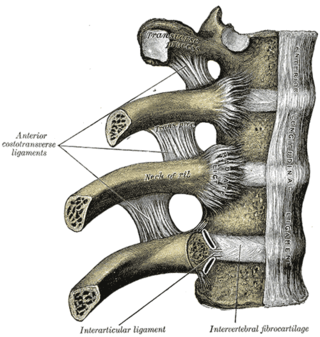
The radiate ligament of head of rib is a ligament of the costovertebral joint that typically connects the anterior edge of the head of each rib, and the side of the bodies of two adjacent vertebrae and their intervertebral discs. The ligament is formed as a thickening of the anterior portion of the joint capsule of the costovertebral joint, and thus reinforces it anteriorly.
The intra-articular ligament of head of rib is a ligament of the articulation of head of rib situated within the joint capsule. It takes the shape of a short, flat band. It joins the crest of head of rib, and the intervertebral disc. It is absent in ribs I and X-XII.
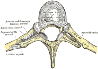
The costotransverse joint is the joint formed between the facet of the tubercle of the rib and the adjacent transverse process of a thoracic vertebra. The costotransverse joint is a plane type of synovial joint which, under physiological conditions, allows only gliding movement.
A costotransverse ligament is ligament of the costotransverse joint which attaches at the neck of a rib, and at the transverse process of its corresponding vertebra. It extends posteriorly from the rib to the vertebra.
A superior costotransverse ligament is a ligament of the costotransverse joint that attaches onto the crest of the neck of a rib, and onto the transverse process of the vertebra superior to the rib.

The pelvis is the lower part of an anatomical trunk, between the abdomen and the thighs, together with its embedded skeleton.

Each vertebra is an irregular bone with a complex structure composed of bone and some hyaline cartilage, that make up the vertebral column or spine, of vertebrates. The proportions of the vertebrae differ according to their spinal segment and the particular species.
References
- 1 2 3 Standring, Susan (2020). Gray's Anatomy: The Anatomical Basis of Clinical Practice (42th ed.). New York. p. 581. ISBN 978-0-7020-7707-4. OCLC 1201341621.
{{cite book}}: CS1 maint: location missing publisher (link) - ↑ Moore, Keith L.; Dalley, Arthur F.; Agur, Anne M. R. (2018). Clinically Oriented Anatomy (8th ed.). Wolters Kluwer. p. 298. ISBN 978-1-4963-4721-3.