
Radiology is the medical specialty that uses medical imaging to diagnose diseases and guide treatment within the bodies of humans and other animals. It began with radiography, but today it includes all imaging modalities. This includes technologies that use no ionizing electromagnetic radiation, such as ultrasonography and magnetic resonance imaging), as well as others that do use radiation, such as computed tomography (CT), fluoroscopy, and nuclear medicine including positron emission tomography (PET). Interventional radiology is the performance of usually minimally invasive medical procedures with the guidance of imaging technologies such as those mentioned above.
Spondyloarthritis (SpA), also known as spondyloarthropathy, is a collection of clinical syndromes that are connected by genetic predisposition and clinical manifestations. The best-known clinical subtypes are enteropathic arthritis (EA), psoriatic arthritis (PsA), ankylosing spondylitis (AS), and reactive arthritis (ReA). Spondyloarthritis typically presents with inflammatory back pain and asymmetrical arthritis, primarily affecting the lower limbs, and enthesitis, inflammation at bone-adhering ligaments, tendons, or joint capsules.
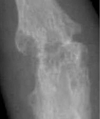
Acute septic arthritis, infectious arthritis, suppurative arthritis, pyogenic arthritis, osteomyelitis, or joint infection is the invasion of a joint by an infectious agent resulting in joint inflammation. Generally speaking, symptoms typically include redness, heat and pain in a single joint associated with a decreased ability to move the joint. Onset is usually rapid. Other symptoms may include fever, weakness and headache. Occasionally, more than one joint may be involved, especially in neonates, younger children and immunocompromised individuals. In neonates, infants during the first year of life, and toddlers, the signs and symptoms of septic arthritis can be deceptive and mimic other infectious and non-infectious disorders.
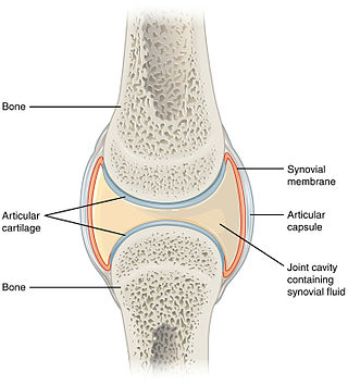
A synovial joint, also known as diarthrosis, joins bones or cartilage with a fibrous joint capsule that is continuous with the periosteum of the joined bones, constitutes the outer boundary of a synovial cavity, and surrounds the bones' articulating surfaces. This joint unites long bones and permits free bone movement and greater mobility. The synovial cavity/joint is filled with synovial fluid. The joint capsule is made up of an outer layer of fibrous membrane, which keeps the bones together structurally, and an inner layer, the synovial membrane, which seals in the synovial fluid.
Dysbaric osteonecrosis or DON is a form of avascular necrosis where there is death of a portion of the bone that is thought to be caused by nitrogen (N2) embolism (blockage of the blood vessels by a bubble of nitrogen coming out of solution) in divers. Although the definitive pathologic process is poorly understood, there are several hypotheses:

A limp is a type of asymmetric abnormality of the gait. Limping may be caused by pain, weakness, neuromuscular imbalance, or a skeletal deformity. The most common underlying cause of a painful limp is physical trauma; however, in the absence of trauma, other serious causes, such as septic arthritis or slipped capital femoral epiphysis, may be present. The diagnostic approach involves ruling out potentially serious causes via the use of X-rays, blood tests, and sometimes joint aspiration. Initial treatment involves pain management. A limp is the presenting problem in about 4% of children who visit hospital emergency departments.

Ankylosis is a stiffness of a joint due to abnormal adhesion and rigidity of the bones of the joint, which may be the result of injury or disease. The rigidity may be complete or partial and may be due to inflammation of the tendinous or muscular structures outside the joint or of the tissues of the joint itself.

A chest radiograph, chest X-ray (CXR), or chest film is a projection radiograph of the chest used to diagnose conditions affecting the chest, its contents, and nearby structures. Chest radiographs are the most common film taken in medicine.
SAPHO syndrome includes a variety of inflammatory bone disorders that may be associated with skin changes. These diseases share some clinical, radiologic, and pathologic characteristics.
An arthropathy is a disease of a joint.

A bone scan or bone scintigraphy is a nuclear medicine imaging technique used to help diagnose and assess different bone diseases. These include cancer of the bone or metastasis, location of bone inflammation and fractures, and bone infection (osteomyelitis).

Transient synovitis of hip is a self-limiting condition in which there is an inflammation of the inner lining of the capsule of the hip joint. The term irritable hip refers to the syndrome of acute hip pain, joint stiffness, limp or non-weightbearing, indicative of an underlying condition such as transient synovitis or orthopedic infections. In everyday clinical practice however, irritable hip is commonly used as a synonym for transient synovitis. It should not be confused with sciatica, a condition describing hip and lower back pain much more common to adults than transient synovitis but with similar signs and symptoms.
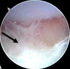
Synovectomy is the surgical removal of the synovial tissue surrounding a joint. This procedure is typically recommended to provide relief from a condition in which the synovial membrane or the joint lining becomes inflamed and irritated and is not controlled by medication alone. If arthritis is not controlled, it can lead to irreversible joint damage. The synovial membrane or "synovium" encloses each joint and also secretes a lubricating fluid that allows different joint motions such as rolling, folding and stretching. When the synovium becomes inflamed or irritated, it increases fluid production, resulting in warmth, tenderness, and swelling in and around the joint.

The fat pad sign, also known as the sail sign, is a potential finding on elbow radiography which suggests a fracture of one or more bones at the elbow. It may indicate an occult fracture that is not directly visible. Its name derives from the fact that it has the shape of a spinnaker (sail). It is caused by displacement of the fat pad around the elbow joint. Both anterior and posterior fat pad signs exist, and both can be found on the same X-ray.
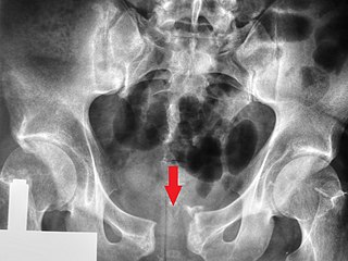
Pubic symphysis diastasis is the separation of normally joined pubic bones, as in the dislocation of the bones, without a fracture that measures radiologically more than 10 mm. Separation of the symphysis pubis is a rare pathology associated with childbirth and has an incidence of 1 in 300 to 1 in 30,000 births. It is usually noticed after delivery but can be observed up to six months postpartum. Risk factors associated with this injury include cephalopelvic disproportion, rapid second stage of labor, epidural anesthesia, severe abduction of the thighs during delivery, or previous trauma to the pelvis. Common signs and symptoms include symphyseal pain aggravated by weight-bearing and walking, a waddling gait, pubic tenderness, and a palpable interpubic gap. Treatment for pubic symphysis diastasis is largely conservative, with treatment modalities including pelvic bracing, bed rest, analgesia, physical therapy, and in some severe cases, surgery.
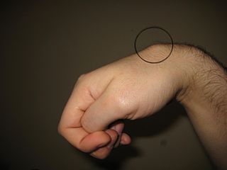
Carpometacarpal bossing is a small, immovable mass of bone on the back of the wrist. The mass occurs in one of the joints between the carpus and metacarpus of the hand, called the carpometacarpal joints, where a small immovable protuberance occurs when this joint becomes swollen or bossed.

Orthopedic pathology, also known as bone pathology is a subspecialty of surgical pathology which deals with the diagnosis and feature of many bone diseases, specifically studying the cause and effects of disorders of the musculoskeletal system. It uses gross and microscopic findings along with the findings of in vivo radiological studies, and occasionally, specimen radiographs to diagnose diseases of the bones.
Protrusio acetabuli is an uncommon defect of the acetabulum, the socket that receives the femoral head to make the hip joint. The hip bone of the pelvic bone/girdle is composed of three bones, the ilium, the ischium and the pubis. In protrusio deformity, there is medial displacement of the femoral head in that the medial aspect of the femoral cortex is medial to the ilioischial line. The socket is too deep and may protrude into the pelvis.

Little League elbow, technically termed medial epicondyle apophysitis, is a condition that is caused by repetitive overhand throwing motions in children. "Little Leaguer's elbow" was coined by Brogdon and Crow in an eponymous 1960 article in the American Journal of Radiology.
Mario Maas is a Dutch professor of radiology, in particular musculoskeletal radiology at the University of Amsterdam and the Academic Medical Centre of Amsterdam.