Ligament (bivalve)

Last updated
This interior view of the hinge line of a blue mussel, Mytilidae shows the external ligament, which is dried out and cracked in this specimen Mytilidae hinge.jpg
This interior view of the hinge line of a blue mussel, Mytilidae shows the external ligament, which is dried out and cracked in this specimen
This interior view of the hinge line of a scallop shell Pectinidae shows the internal ligament, located in the resilifer. Pectinidae hinge.jpg
This interior view of the hinge line of a scallop shell Pectinidae shows the internal ligament, located in the resilifer.
Interior view of the hinge ligament of Tridacna derasa Tridacna derasa hinge ligament.jpg
Interior view of the hinge ligament of Tridacna derasa

A hinge ligament is a crucial part of the anatomical structure of a bivalve shell, i.e. the shell of a bivalve mollusk. The shell of a bivalve has two valves and these are joined by the ligament at the dorsal edge of the shell. The ligament is made of a strong, flexible and elastic, fibrous, proteinaceous material which is usually pale brown, dark brown or black in color.

Contents

In life, the shell needs to be able to open a little (to allow the foot and siphons to protrude) and then close again. As well as connecting the two bivalve shells together at the hinge line, the ligament also functions as a spring which automatically opens the valves when the adductor muscle or muscles (that close the valves) relax.

Composition

The ligament is an uncalcified elastic structure comprised in its most minimal state of two layers: a lamellar layer and a fibrous layer. The lamellar layer consists entirely of organic material (a protein and collagen matrix), is generally brown in color, and is elastic in response to both compressional and tensional stresses. The fibrous layer is made of aragonite fibers and organic material, is lighter in color and often iridescent, and is elastic only under compressional stress. [1] The protein responsible for the elasticity of the ligament is abductin, which has enormous elastic resiliency: this resiliency is what causes the valves of the bivalve mollusk to open when the adductor muscles relax. [2]

Ligaments that are simple morphologically have a central fibrous layer between the anterior and posterior lamellar layers. Repetitive ligaments are morphologically more complex, and display additional, repeated layers. [3] A recent study using scanning electron microscopy(SEM), X-ray diffraction (XRD), and infrared spectroscopy (FTIR), found that some bivalve mollusks have a third type of fibrous layer in the ligament (located in the middle) which has a unique spring-like protein fiber (ca. 120 nm in diameter) structure, stretching continuously from the left to right valve. [4]

Elastic opening of valves

When the adductor muscles of a bivalve mollusk contract, the valves close, which compresses the ligament. When the adductor muscles relax again, the elastic resiliency of the ligament reopens the shell. Scallops (Pectinidae) swim through the water column by rapidly and repeatedly clapping (opening and closing) their valves. An interesting fact about scallops swimming in this manner is that they recover a greater percentage of the work (as defined by physics) performed through the elasticity of their abductin than do other bivalves (which are more sedentary clams). [2]

Taxonomic use

The hinge ligament of a bivalve shell can be either internal, external, or both, and is an example of complex development. [5] Various types of hinge ligaments have been found in living species (i.e. extant species), and the ligaments can be reconstructed in most fossil bivalves based on their sites of attachment on the shell. The taxonomic distribution of ligament types among families of bivalves has been used by paleontologists and malacologists as a means of inferring phylogenic evolution. [1]

Hinge ligament types

External hinge ligaments may be described as having an "orientation" that is amphidetic (between the beaks), opisthodetic (behind/ posterior to the beaks), or, rarely, prosodetic (before the beaks). Then, there are four main "structural types": alivincular (a flattened, usually triangular area with a central fibrous layer and a peripheral lamellar layer), duplivincular (alternating bands of fibrous and lamellar layers forming chevrons on the cardinal area), parivincular (a single arched structure behind the beaks), and planivincular (a long ligament with a slight arch that extends behind the beaks). [6]

An internal ligament is usually called a resilium and is attached to a resilifer or chrondophore, which is a depression or pit inside the shell near the umbo. [5] [7]

Related Research Articles

<span class="mw-page-title-main">Bivalvia</span> Class of molluscs

Bivalvia or bivalves, in previous centuries referred to as the Lamellibranchiata and Pelecypoda, is a class of aquatic molluscs that have laterally compressed soft bodies enclosed by a calcified exoskeleton consisting of a hinged pair of half-shells known as valves. As a group, bivalves have no head and lack some typical molluscan organs such as the radula and the odontophore. Their gills have evolved into ctenidia, specialised organs for feeding and breathing.
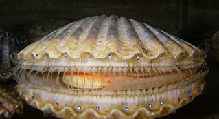
<span class="mw-page-title-main">Scallop</span> Common name for several shellfish, many edible

Scallop is a common name that encompasses various species of marine bivalve mollusks in the taxonomic family Pectinidae, the scallops. However, the common name "scallop" is also sometimes applied to species in other closely related families within the superfamily Pectinoidea, which also includes the thorny oysters.

<span class="mw-page-title-main">Arcida</span> Order of molluscs

The Arcida is an extant order of bivalve molluscs. This order dates back to the lower Ordovician period. They are distinguished from related groups, such as the mussels, by having a straight hinge to the shells, and the adductor muscles being of equal size. The duplivincular ligament, taxodont dentition, and a shell microstructure consisting of the outer crossed lamellar and inner complex crossed lamellar layers are defining characters of this order.
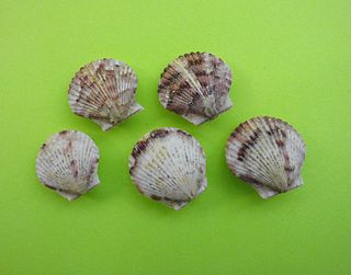
<i>Argopecten gibbus</i> Species of bivalve

Argopecten gibbus, the Atlantic calico scallop, is a species of medium-sized edible marine bivalve mollusk in the family Pectinidae, the scallops.

Freshwater bivalves are molluscs of the order Bivalvia that inhabit freshwater ecosystems. They are one of the two main groups of freshwater molluscs, along with freshwater snails.

<span class="mw-page-title-main">Bivalve shell</span> Seashell

A bivalve shell is the enveloping exoskeleton or shell of a bivalve mollusc, composed of two hinged halves or valves. The two half-shells, called the "right valve" and "left valve", are joined by a ligament and usually articulate with one another using structures known as "teeth" which are situated along the hinge line. In many bivalve shells, the two valves are symmetrical along the hinge line — when truly symmetrical, such an animal is said to be equivalved; if the valves vary from each other in size or shape, inequivalved. If symmetrical front-to-back, the valves are said to be equilateral, and are otherwise considered inequilateral.

<span class="mw-page-title-main">Mollusc shell</span> Exoskeleton of an animal in the phylum Mollusca

The molluscshell is typically a calcareous exoskeleton which encloses, supports and protects the soft parts of an animal in the phylum Mollusca, which includes snails, clams, tusk shells, and several other classes. Not all shelled molluscs live in the sea; many live on the land and in freshwater.

<i>Fordilla</i> Extinct genus of bivalves

Fordilla is an extinct genus of early bivalves, one of two genera in the extinct family Fordillidae. The genus is known solely from Early Cambrian fossils found in North America, Greenland, Europe, the Middle East, and Asia. The genus currently contains three described species, Fordilla germanica, Fordilla sibirica, and the type species Fordilla troyensis.

Pojetaia is an extinct genus of early bivalves, one of two genera in the extinct family Fordillidae. The genus is known solely from Early to Middle Cambrian fossils found in North America, Greenland, Europe, North Africa, Asia, and Australia. The genus currently contains two accepted species, Pojetaia runnegari, the type species, and Pojetaia sarhroensis, though up to seven species have been proposed. The genera Buluniella, Jellia, and Oryzoconcha are all considered synonyms of Pojetaia.

<span class="mw-page-title-main">Fordillidae</span> Extinct family of bivalves

Fordillidae is an extinct family of early bivalves and one of two families in the extinct superfamily Fordilloidea. The family is known from fossils of early to middle Cambrian age found in North America, Greenland, Europe, the Middle East, Asia, and Australia. The family currently contains two genera, Fordilla and Pojetaia, each with up to three described species. Due to the size and age of the fossil specimens, Fordillidae species are included as part of the Turkish Small shelly fauna.

<span class="mw-page-title-main">Fordilloidea</span> Extinct superfamily of bivalves

Fordilloidea is an extinct superfamily of early bivalves containing two described families, Fordillidae and Camyidae and the only superfamily in the order Fordillida. The superfamily is known from fossils of early to middle Cambrian age found in North America, Greenland, Europe, the Middle East, Asia, and Australia. Fordillidae currently contains two genera, Fordilla and Pojetaia each with up to three described species while Camyidae only contains a single genus Camya with one described species, Camya asy. Due to the size and age of the fossil specimens, Fordillidae species are included as part of the Turkish Small shelly fauna.
<span class="mw-page-title-main">Antarctic scallop</span> Genus of bivalves

The Antarctic scallop is a species of bivalve mollusc in the large family of scallops, the Pectinidae. It was thought to be the only species in the genus Adamussium until an extinct Pliocene species was described in 2016. Its exact relationship to other members of the Pectinidae is unclear. It is found in the ice-cold seas surrounding Antarctica, sometimes at great depths.

<i>Cucullaea labiata</i> Species of bivalve

Cucullaea labiata is a species of saltwater clam or ark shell, a marine bivalve mollusk in the family Cucullaeidae.

<span class="mw-page-title-main">Hinge teeth</span>

Hinge teeth are part of the anatomical structure of the inner surface of a bivalve shell, i.e. the shell of a bivalve mollusk. Bivalves by definition have two valves, which are joined together by a strong and flexible ligament situated on the hinge line at the dorsal edge of the shell. In life, the shell needs to be able to open slightly to allow the foot and siphons to protrude, and then close again, without the valves moving out of alignment with one another. To make this possible, in most cases the two valves are articulated using an arrangement of structures known as hinge teeth. Like the ligament, the hinge teeth are also situated along the hinge line of the shell, in most cases.

<span class="mw-page-title-main">Resilifer</span>

A resilifer is a part of the shell of certain bivalve mollusks. It is either a recess or a process, the function of which is the attachment of an internal ligament, which holds the two valves together.

<span class="mw-page-title-main">Abductin</span>

Abductin is a naturally occurring elastomeric protein found in the hinge ligament of bivalve mollusks. It is unique as it is the only natural elastomer with compressible elasticity, as compared to resilin, spider silk, and elastin. Its name was proposed from the fact that it functions as the abductor of the valves of bivalve mollusks.

<span class="mw-page-title-main">Pallial line</span>

The pallial line is a mark on the interior of each valve of the shell of a bivalve mollusk. This line shows where all of the mantle muscles were attached in life. In clams with two adductor muscles the pallial line usually joins the marks known as adductor muscle scars, which are where the adductor muscles attach.

<span class="mw-page-title-main">Adductor muscles (bivalve)</span> Main muscular system in bivalves

The adductor muscles are the main muscular system in bivalve mollusks. In many parts of the world, when people eat scallops, the adductor muscles are the only part of the animal which is eaten. Adductor muscles leave noticeable scars or marks on the interior of the shell's valves. Those marks are often used by scientists who are in the process of identifying empty shells to determine their correct taxonomic placement.

<span class="mw-page-title-main">Resilium</span>

In anatomy, a resilium is part of the shell of certain bivalve mollusks. It is an internal ligament, which holds the two valves together and is located in a pit or depression known as the resilifer.

<span class="mw-page-title-main">Ostreoidea</span> Superfamily of bivalves

Ostreoidea is a taxonomic superfamily of bivalve marine mollusc, sometimes simply identified as oysters, containing two families. The ostreoids are characterized in part by the presence of a well developed axial rod. Anal flaps are known to exist within the family Ostreidae but not within the more-primitive Gryphaeidae. The scar from the adductor muscle is simple, with a single, central scar. In the majority, the right valve is less convex than the left.

References

  1. 1 2 A Theoretical Morphologic Analysis of Bivalve Ligaments, Takao Ubukata, Paleobiology Vol. 29, No. 3 (Summer, 2003), pp. 369-380
  2. 1 2 Steven Vogel (2003) Comparative Biomechanics: Life's Physical World. Princeton: Princeton University Press. 580 p. at p. 304
  3. Morphology and postlarval development of the ligament of Thracia phaseolina (Bivalvia: Thraciidae), with a discussion of model choice in allometric studies, André F. Sartori1 and Alexander D. Ball, J. Mollus. Stud. (2009) 75(3):295-304
  4. A new structural model of bivalve ligament from Solen grandis. Zengqiong H, Gangsheng Z., Micron. 2011 Oct; 42(7):706-11
  5. 1 2 "Evolution on the half shell – Assembling the Tree of Life: the Bivalve Mollusks". 2012-08-26. Archived from the original on 2012-08-26. Retrieved 2023-08-18.
  6. "Marine bivalve shells of the British Isles - An introduction to shell structures". National Museum of Wales. 2014. Archived from the original on 23 February 2015. Retrieved 22 October 2014.
  7. Huber, Markus (2010). Compendium of Bivalves. A Full-color Guide to 3'300 of the World's Marine Bivalves. A Status on Bivalvia after 250 Years of Research. Hackenheim: ConchBooks. pp. 901 pp. + CD. ISBN   978-3-939767-28-2, at p. 59

General references

E.R. Trueman, General features of Bivalvia. In: Moore R.C., editor. Bivalvia. Ligament. In: Treatise on invertebrate paleontology. Vol. 2. Geological Society of America and University of Kansas Press; 1969. p. N58-N64. Part N - Mollusca, Bivalvia Vol. 6.

T.R. Waller, The evolution of ligament systems in the Bivalvia. In: Morton B., editor. Proceedings of a Memorial Symposium in Honour of Sir Charles Maurice Yonge, Edinburgh, 1986. Hong Kong: Hong Kong University Press; 1990. p. 49-71.

J. G. Carter, Evolutionary significance of shell microstructure in the Paleotaxodonta, Pteriomorphia and Isofilibranchia (Bivalvia: Mollusca). In: Carter J.G., editor. Skeletal biomineralization: patterns, processes, and evolutionary trends. New York: Van Nostrand Reinhold; 1990. p. 135-296.