
Histology, also known as microscopic anatomy or microanatomy, is the branch of biology that studies the microscopic anatomy of biological tissues. Histology is the microscopic counterpart to gross anatomy, which looks at larger structures visible without a microscope. Although one may divide microscopic anatomy into organology, the study of organs, histology, the study of tissues, and cytology, the study of cells, modern usage places all of these topics under the field of histology. In medicine, histopathology is the branch of histology that includes the microscopic identification and study of diseased tissue. In the field of paleontology, the term paleohistology refers to the histology of fossil organisms.

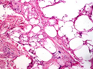
A lipoma is a benign tumor made of fat tissue. They are generally soft to the touch, movable, and painless. They usually occur just under the skin, but occasionally may be deeper. Most are less than 5 cm (2.0 in) in size. Common locations include upper back, shoulders, and abdomen. It is possible to have several lipomas.

Staining is a technique used to enhance contrast in samples, generally at the microscopic level. Stains and dyes are frequently used in histology, in cytology, and in the medical fields of histopathology, hematology, and cytopathology that focus on the study and diagnoses of diseases at the microscopic level. Stains may be used to define biological tissues, cell populations, or organelles within individual cells.

Leydig cells, also known as interstitial cells of the testes and interstitial cells of Leydig, are found adjacent to the seminiferous tubules in the testicle and produce testosterone in the presence of luteinizing hormone (LH). They are polyhedral in shape and have a large, prominent nucleus, an eosinophilic cytoplasm, and numerous lipid-filled vesicles.

Adipocytes, also known as lipocytes and fat cells, are the cells that primarily compose adipose tissue, specialized in storing energy as fat. Adipocytes are derived from mesenchymal stem cells which give rise to adipocytes through adipogenesis. In cell culture, adipocyte progenitors can also form osteoblasts, myocytes and other cell types.

Histopathology is the microscopic examination of tissue in order to study the manifestations of disease. Specifically, in clinical medicine, histopathology refers to the examination of a biopsy or surgical specimen by a pathologist, after the specimen has been processed and histological sections have been placed onto glass slides. In contrast, cytopathology examines free cells or tissue micro-fragments.

Eosinophilic is the staining of tissues, cells, or organelles after they have been washed with eosin, a dye.

Liposarcomas are the most common subtype of soft tissue sarcomas, accounting for at least 20% of all sarcomas in adults. Soft tissue sarcomas are rare neoplasms with over 150 different histological subtypes or forms. Liposarcomas arise from the precursor lipoblasts of the adipocytes in adipose tissues. Adipose tissues are distributed throughout the body, including such sites as the deep and more superficial layers of subcutaneous tissues as well as in less surgically accessible sites like the retroperitoneum and visceral fat inside the abdominal cavity.

Papanicolaou stain is a multichromatic (multicolored) cytological staining technique developed by George Papanicolaou in 1942. The Papanicolaou stain is one of the most widely used stains in cytology, where it is used to aid pathologists in making a diagnosis. Although most notable for its use in the detection of cervical cancer in the Pap test or Pap smear, it is also used to stain non-gynecological specimen preparations from a variety of bodily secretions and from small needle biopsies of organs and tissues. Papanicolaou published three formulations of this stain in 1942, 1954, and 1960.
Hematoxylin and eosin stain is one of the principal tissue stains used in histology. It is the most widely used stain in medical diagnosis and is often the gold standard. For example, when a pathologist looks at a biopsy of a suspected cancer, the histological section is likely to be stained with H&E.

3T3-L1 is a sub clonal cell line derived from the original 3T3 Swiss albino cell line of 1962. The 3T3 original cell line was isolated from a mouse embryo and propagated for this specific line of 3T3 cells is used to study adipose tissue-related diseases and dysfunctions. The 3T3-L1 Swiss subclone line has been widely utilized, since its development, due to its affinity for lipid droplet deposition in vitro. 3T3-L1 cells have a fibroblast-like morphology, but, under appropriate conditions, the cells differentiate into an adipocyte-like phenotype, providing an exemplar model for white adipocytes. 3T3-L1 cells can be utilized to study a number of cellular and molecular mechanisms related to insulin-resistance, obesity, and diabetes in vitro. Aside from its usages, this cell line is widely developed and can be purchased for continuous propagation for numerous research studies. 3T3-L1 cells of the adipocyte morphology increase the synthesis and accumulation of triglycerides and acquire the signet ring appearance of adipose cells. These cells are also sensitive to lipogenic and lipolytic hormones, as well as drugs, including epinephrine, isoproterenol, and insulin.

Germ cell neoplasia in situ (GCNIS) represents the precursor lesion for many types of testicular germ cell tumors.
A hibernoma is a benign neoplasm of vestigial brown fat. They were first described under the name ‘pseudolipoma’ by the German physician H. Merkel in 1906 and the term hibernoma was proposed by the French anatomist Louis Gery in 1914 because of its resemblance to brown fat in hibernating animals.
Benign lipoblastomatosis is a tumor consisting of fetal-embryonal adipocytes, frequently confused with a liposarcoma, affecting exclusively infants and young children, with approximately 90% of cases occurring before 3 years of age. The term lipoblastomatosis was first used by Vellios et al. in 1958, at which point the tumor became generally accepted as a distinctive entity. Today Diffuse lipoblastoma is the preferred term for lipoblastomatosis. The tumor is rare, accounting for less than 1% of all childhood neoplasm, and it has been found to be more common in males than females. It often presents as an asymptomatic rapidly enlarging mass, occurring more often in the soft tissues of the extremities.
Chondroid lipomas are deep-seated, firm, yellow tumors that characteristically occur on the legs of women. They exhibit a characteristic genetic translocation t(11;16) with a resulting C11orf95-MKL2 fusion oncogene.
Pleomorphic lipomas, like spindle-cell lipomas, occur for the most part on the backs and necks of elderly men, and are characterized by floret giant cells with overlapping nuclei.

In histopathology, ballooning degeneration, formally ballooning degeneration of hepatocytes, is a form of liver parenchymal cell death.

Adipogenesis is the formation of adipocytes from stem cells. It involves 2 phases, determination, and terminal differentiation. Determination is mesenchymal stem cells committing to the adipocyte precursor cells, also known as lipoblasts or preadipocytes which lose the potential to differentiate to other types of cells such as chondrocytes, myocytes, and osteoblasts. Terminal differentiation is that preadipocytes differentiate into mature adipocytes. Adipocytes can arise either from preadipocytes resident in adipose tissue, or from bone-marrow derived progenitor cells that migrate to adipose tissue.
A rhabdomyoblast is a cell type which is found in some rhabdomyosarcomas. When found histologically, a rhabdomyoblast aids the diagnosis of embryonal, alveolar, spindle cell/sclerosing, and pleomorphic rhabdomyosarcomas; however, in a tumor, expression of the rhabdomyoblast phenotype is not the only factor in diagnosing a rhabdomyosarcoma. Mesenchymal malignancies can exhibit this phenotype as well. Immunohistochemistry techniques allow for the sensitive detection of desmin, vimentin, muscle specific actin, and MyoD1. Similarly the rhabdomyoblast phenotype can be detected morphologically. Rhabdomyoblasts are early stage mesenchymal cells, having the potential to differentiate into a wide range of skeletal cells. Each stage of differentiation exhibits unique and distinguishable histological characteristics. In its initial from, stellate cells with amphiphilic cytoplasm and ovular central nuclei are observed. Commonly referred to as rhabdoid features, the maturing rhabdomyoblast will likely exhibit low levels of eosinophilic cytoplasm in proximal distances to the nucleus. As maturation and differentiation progress, the cell's cytoplasmic levels of white blood cells increase; additionally, elongated shapes, commonly depicted as “tadpole”, “strap” and "spider cells", are observed. In the concluding phase of differentiation, the white blood cell rich cytoplasm appears bright and exhibits cross-striation. The highly regulated organization of actin and myosin microfilaments in contractile proteins results in this appearance.
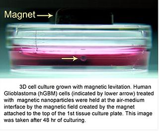
3D cell culturing by Magnetic LevitationMethod (MLM) is the application of growing 3D tissue by inducing cells treated with magnetic nanoparticle assemblies in spatially varying magnetic fields, using neodymium magnetic drivers and promoting cell-to-cell interactions by levitating the cells up to the air/liquid interface of a standard petri dish. The magnetic nanoparticle assemblies consist of magnetic iron oxide nanoparticles, gold nanoparticles, and the polymer polylysine. 3D cell culturing is scalable, with the capability of culturing as few as 500 cells up to millions of cells, or from a single dish to high-throughput low volume systems. Once magnetized cultures are generated, they can also be used as the building block material, or the "ink" for the magnetic 3D bioprinting process.

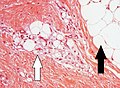 Lipoblasts (white arrow) and lipocytes (black arrow), in a case of lipoblastoma
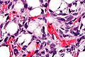
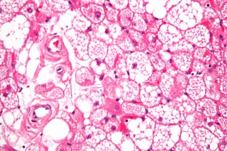
 Histopathology of liposarcoma, H&E stain, with the main features: [5]
 Lipid-laden histiocytes may mimic lipoblasts, but have lightly eosinophilic cytoplasm and a small normochromatic nuclei which are not hollowed out from the lipid vacuoles. [6]


