
A lipoma is a benign tumor made of fat tissue. They are generally soft to the touch, movable, and painless. They usually occur just under the skin, but occasionally may be deeper. Most are less than 5 cm (2.0 in) in size. Common locations include upper back, shoulders, and abdomen. It is possible to have a number of lipomas.

Adipocytes, also known as lipocytes and fat cells, are the cells that primarily compose adipose tissue, specialized in storing energy as fat. Adipocytes are derived from mesenchymal stem cells which give rise to adipocytes through adipogenesis. In cell culture, adipocyte progenitors can also form osteoblasts, myocytes and other cell types.

A myxoma is a myxoid tumor of primitive connective tissue. It is most commonly found in the heart but can also occur in other locations.

A benign tumor is a mass of cells (tumor) that does not invade neighboring tissue or metastasize. Compared to malignant (cancerous) tumors, benign tumors generally have a slower growth rate. Benign tumors have relatively well differentiated cells. They are often surrounded by an outer surface or stay contained within the epithelium. Common examples of benign tumors include moles and uterine fibroids.

Liposarcomas are the most common subtype of soft tissue sarcomas, accounting for at least 20% of all sarcomas in adults. Soft tissue sarcomas are rare neoplasms with over 150 different histological subtypes or forms. Liposarcomas arise from the precursor lipoblasts of the adipocytes in adipose tissues. Adipose tissues are distributed throughout the body, including such sites as the deep and more superficial layers of subcutaneous tissues as well as in less surgically accessible sites like the retroperitoneum and visceral fat inside the abdominal cavity.

An ameloblastic fibroma is a fibroma of the ameloblastic tissue, that is, an odontogenic tumor arising from the enamel organ or dental lamina. It may be either truly neoplastic or merely hamartomatous. In neoplastic cases, it may be labeled an ameloblastic fibrosarcoma in accord with the terminological distinction that reserves the word fibroma for benign tumors and assigns the word fibrosarcoma to malignant ones. It is more common in the first and second decades of life, when odontogenesis is ongoing, than in later decades. In 50% of cases an unerupted tooth is involved.
Angiomyxoma is a myxoid tumor involving the blood vessels.
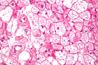
A hibernoma is a benign neoplasm of vestigial brown fat. The term was originally used by the French anatomist Louis Gery in 1914.
Fibrous hamartoma of infancy (FHI) is a rare, typically painless, benign tumor that develops in the subcutaneous tissues of the axilla, arms, external genitalia, or, less commonly, various other areas. It is diagnosed in children who are usually less than 2 years old or, in up to 20% of cases, develops in utero and is diagnosed in an infant at birth.
Benign lipoblastomatosis is a tumor consisting of fetal-embryonal adipocytes, frequently confused with a liposarcoma, affecting exclusively infants and young children, with approximately 90% of cases occurring before 3 years of age. The term lipoblastomatosis was first used by Vellios et al. in 1958, at which point the tumor became generally accepted as a distinctive entity. Today Diffuse lipoblastoma is the preferred term for lipoblastomatosis. The tumor is rare, accounting for less than 1% of all childhood neoplasm, and it has been found to be more common in males than females. It often presents as an asymptomatic rapidly enlarging mass, occurring more often in the soft tissues of the extremities.

A Neurothekeoma (NT) is a type of rare benign cutaneous tumor that usually develops on the head and neck. They often occur in the second and early third decades of life and tend to afflict women more frequently than men. First described by Gallager and Helwig, who proposed the term in order to reflect the presumed origin of the lesion from nerve sheath. Microscopically, the lesions described closely resembled the tumor, "nerve sheath myxoma (NSM)", an entity first described by Harkin and Reed. The latter had, through the years, been variously described as "bizarre cutaneous neurofibroma", "myxoma of nerve sheath", and "pacinian neurofibroma".
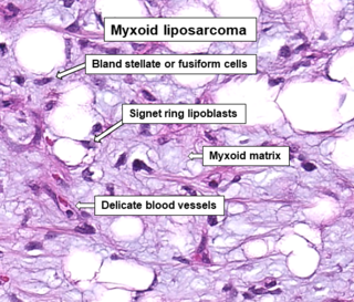
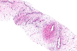
A myxoid liposarcoma is a malignant adipose tissue neoplasm of myxoid appearance histologically.

A lipoblast is a precursor cell for an adipocyte.

Low-grade fibromyxoid sarcoma (LGFMS) is a rare type of low-grade sarcoma first described by H. L. Evans in 1987. LGFMS are soft tissue tumors of the mesenchyme-derived connective tissues; on microscopic examination, they are found to be composed of spindle-shaped cells that resemble fibroblasts. These fibroblastic, spindle-shaped cells are neoplastic cells that in most cases of LGFMS express fusion genes, i.e. genes composed of parts of two different genes that form as a result of mutations. The World Health Organization (2020) classified LGFMS as a specific type of tumor in the category of malignant fibroblastic and myofibroblastic tumors.

Mammary-type myofibroblastoma (MFB), also named mammary and extramammary myofibroblastoma, was first termed myofibrolastoma of the breast, or, more simply, either mammary myofibroblastoma (MMFB) or just myofibroblastoma. The change in this terminology occurred because the initial 1987 study and many subsequent studies found this tumor only in breast tissue. However, a 2001 study followed by numerous reports found tumors with the microscopic histopathology and other key features of mammary MFB in a wide range of organs and tissues. Further complicating the issue, early studies on MFB classified it as one of various types of spindle cell tumors that, except for MFB, were ill-defined. These other tumors, which have often been named interchangeably in different reports, are: myelofibroblastoma, benign spindle cell tumor, fibroma, spindle cell lipoma, myogenic stromal tumor, and solitary stromal tumor. Finally, studies suggest that spindle cell lipoma and cellular angiofibroma are variants of MFB. Here, the latter two tumors are tentatively classified as MFB variants but otherwise MFB is described as it is more strictly defined in most recent publications. The World Health Organization in 2020 classified mammary type myofibroblastoma tumors and myofibroblastoma tumors as separate tumor forms within the category of fibroblastic and myofibroblastic tumors.

Familial multiple lipomatosis is a hereditary adipose tissue disorder that is characterized by the formation of multiple lipomas that occur in a particular distribution. The lipomas are well-encapsulated, slow-growing, benign fatty tumors. The distribution is defined as being focused in the trunk of the body and extremities. Familial Multiple Lipomatosis can be identified when multiple lipomas occur in multiple family members that span different generations. Some people may have hundreds of lipomas present.
Lipofibromatosis (LPF) is an extremely rare soft tissue tumor which was first clearly described in 2000 by Fetsch et al as a strictly pediatric, locally invasive, and often recurrent tumor. It is nonetheless a non-metastasizing, i.e. benign, tumor. While even the more recent literature has sometimes regarded LPF as a strictly childhood disorder, rare cases of LPF has been diagnosed in adults. The diagnosis of lipofibromatosis should not be automatically discarded because of an individual's age.
Myxofibrosarcoma (MFS), although a rare type of tumor, is one of the most common soft tissue sarcomas, i.e. cancerous tumors, that develop in the soft tissues of elderly individuals. Initially considered to be a type of histiocytoma termed fibrous histiocytoma or myxoid variant of malignant fibrous histiocytoma, Angervall et al. termed this tumor myxofibrosarcoma in 1977. In 2020, the World Health Organization reclassified MFS as a separate and distinct tumor in the category of malignant fibroblastic and myofibroblastic tumors.
Cellular angiofibroma (CAF) is a rare, benign tumor of superficial soft tissues that was first described by M. R. Nucci et al. in 1997. These tumors occur predominantly in the distal parts of the female and male reproductive systems, i.e. in the vulva-vaginal and inguinal-scrotal areas, respectively, or, less commonly, in various other superficial soft tissue areas throughout the body. CAF tumors develop exclusively in adults who typically are more than 30 years old.
Angiofibroma of soft tissue (AFST), also termed angiofibroma, not otherwise specified, is a recently recognized and rare disorder that was classified in the category of benign fibroblastic and myofibroblastic tumors by the World Health Organization in 2020. An AFST tumor is a neoplasm that was first described by A. Mariño-Enríquez and C.D. Fletcher in 2012.

