Related Research Articles

Ian Lancaster Fleming was an English writer, best known for his postwar James Bond series of spy novels. Fleming came from a wealthy family connected to the merchant bank Robert Fleming & Co., and his father was the Member of Parliament (MP) for Henley from 1910 until his death on the Western Front in 1917. Educated at Eton, Sandhurst, and, briefly, the universities of Munich and Geneva, Fleming moved through several jobs before he started writing.

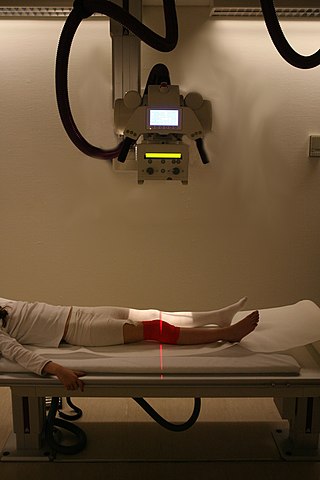
Radiography is an imaging technique using X-rays, gamma rays, or similar ionizing radiation and non-ionizing radiation to view the internal form of an object. Applications of radiography include medical and industrial radiography. Similar techniques are used in airport security,. To create an image in conventional radiography, a beam of X-rays is produced by an X-ray generator and it is projected towards the object. A certain amount of the X-rays or other radiation are absorbed by the object, dependent on the object's density and structural composition. The X-rays that pass through the object are captured behind the object by a detector. The generation of flat two-dimensional images by this technique is called projectional radiography. In computed tomography, an X-ray source and its associated detectors rotate around the subject, which itself moves through the conical X-ray beam produced. Any given point within the subject is crossed from many directions by many different beams at different times. Information regarding the attenuation of these beams is collated and subjected to computation to generate two-dimensional images on three planes which can be further processed to produce a three-dimensional image.

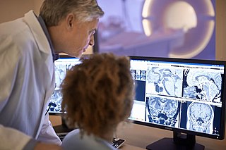
Radiology is the medical specialty that uses medical imaging to diagnose diseases and guide treatment within the bodies of humans and other animals. It began with radiography, but today it includes all imaging modalities. This includes technologies that use no ionizing electromagnetic radiation, such as ultrasonography and magnetic resonance imaging), as well as others that do use radiation, such as computed tomography (CT), fluoroscopy, and nuclear medicine including positron emission tomography (PET). Interventional radiology is the performance of usually minimally invasive medical procedures with the guidance of imaging technologies such as those mentioned above.

An endoscopy is a procedure used in medicine to look inside the body. The endoscopy procedure uses an endoscope to examine the interior of a hollow organ or cavity of the body. Unlike many other medical imaging techniques, endoscopes are inserted directly into the organ.

Fluoroscopy, informally referred to as "fluoro", is an imaging technique that uses X-rays to obtain real-time moving images of the interior of an object. In its primary application of medical imaging, a fluoroscope allows a surgeon to see the internal structure and function of a patient, so that the pumping action of the heart or the motion of swallowing, for example, can be watched. This is useful for both diagnosis and therapy and occurs in general radiology, interventional radiology, and image-guided surgery.

Angiography or arteriography is a medical imaging technique used to visualize the inside, or lumen, of blood vessels and organs of the body, with particular interest in the arteries, veins, and the heart chambers. Modern angiography is performed by injecting a radio-opaque contrast agent into the blood vessel and imaging using X-ray based techniques such as fluoroscopy.
Operation Mincemeat was a successful British deception operation of the Second World War to disguise the 1943 Allied invasion of Sicily. Two members of British intelligence obtained the body of Glyndwr Michael, a tramp who died from eating rat poison, dressed him as an officer of the Royal Marines and placed personal items on him identifying him as the fictitious Captain William Martin. Correspondence between two British generals that suggested that the Allies planned to invade Greece and Sardinia, with Sicily as merely the target of a feint, was also placed on the body.

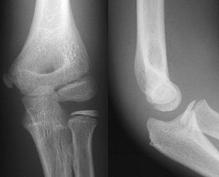
A joint dislocation, also called luxation, occurs when there is an abnormal separation in the joint, where two or more bones meet. A partial dislocation is referred to as a subluxation. Dislocations are often caused by sudden trauma on the joint like an impact or fall. A joint dislocation can cause damage to the surrounding ligaments, tendons, muscles, and nerves. Dislocations can occur in any major joint or minor joint. The most common joint dislocation is a shoulder dislocation.

A chest radiograph, chest X-ray (CXR), or chest film is a projection radiograph of the chest used to diagnose conditions affecting the chest, its contents, and nearby structures. Chest radiographs are the most common film taken in medicine.

A microform is a scaled-down reproduction of a document, typically either photographic film or paper, made for the purposes of transmission, storage, reading, and printing. Microform images are commonly reduced to about 4% or 1⁄25 of the original document size. For special purposes, greater optical reductions may be used.
X-rays are a form of electromagnetic radiation or radiographs: photographs made with X-rays.

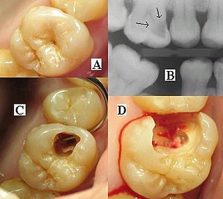
Dental radiographs, commonly known as X-rays, are radiographs used to diagnose hidden dental structures, malignant or benign masses, bone loss, and cavities.

Projectional radiography, also known as conventional radiography, is a form of radiography and medical imaging that produces two-dimensional images by X-ray radiation. The image acquisition is generally performed by radiographers, and the images are often examined by radiologists. Both the procedure and any resultant images are often simply called 'X-ray'. Plain radiography or roentgenography generally refers to projectional radiography. Plain radiography can also refer to radiography without a radiocontrast agent or radiography that generates single static images, as contrasted to fluoroscopy, which are technically also projectional.

An abdominal x-ray is an x-ray of the abdomen. It is sometimes abbreviated to AXR, or KUB.

The program Coot is used to display and manipulate atomic models of macromolecules, typically of proteins or nucleic acids, using 3D computer graphics. It is primarily focused on building and validation of atomic models into three-dimensional electron density maps obtained by X-ray crystallography methods, although it has also been applied to data from electron microscopy.

The European Underwater Federation (EUF) is an umbrella organisation representing the interests of scuba diver training organisations operating in both the not for profit and for profit sectors within Europe.

Flat-panel detectors are a class of solid-state x-ray digital radiography devices similar in principle to the image sensors used in digital photography and video. They are used in both projectional radiography and as an alternative to x-ray image intensifiers (IIs) in fluoroscopy equipment.

Solar wind Magnetosphere Ionosphere Link Explorer (SMILE) is a planned joint venture mission between the European Space Agency and the Chinese Academy of Sciences. SMILE will image for the first time the magnetosphere of the Sun in soft X-rays and UV during up to 40 hours per orbit, improving our understanding of the dynamic interaction between the solar wind and Earth's magnetosphere. The prime science questions of the SMILE mission are
X-ray motion analysis is a technique used to track the movement of objects using X-rays. This is done by placing the subject to be imaged in the center of the X-ray beam and recording the motion using an image intensifier and a high-speed camera, allowing for high quality videos sampled many times per second. Depending on the settings of the X-rays, this technique can visualize specific structures in an object, such as bones or cartilage. X-ray motion analysis can be used to perform gait analysis, analyze joint movement, or record the motion of bones obscured by soft tissue. The ability to measure skeletal motions is a key aspect to one's understanding of vertebrate biomechanics, energetics, and motor control.
Michael Norman George James was Emeritus Distinguished Professor of Biochemistry at the University of Alberta. He was elected a Fellow of the Royal Society (FRS) in 1989 for "substantial contributions to the improvement of natural knowledge". He was appointed to the Order of Canada in June 2023, with the rank of Officer. He lived in Edmonton.
References
- John Macintyre universitystory.gla.ac.uk
- Macintyre's X-Ray Film youtube.com
- JOHN MACINTYRE gdl.cdlr.strath.ac.uk