In optics, an index-matching material is a substance, usually a liquid, cement (adhesive), or gel, which has an index of refraction that closely approximates that of another object.

Time of flight (ToF) is the measurement of the time taken by an object, particle or wave to travel a distance through a medium. This information can then be used to measure velocity or path length, or as a way to learn about the particle or medium's properties. The traveling object may be detected directly or indirectly. Time of flight technology has found valuable applications in the monitoring and characterization of material and biomaterials, hydrogels included.

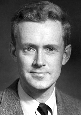
Edward Mills Purcell was an American physicist who shared the 1952 Nobel Prize for Physics for his independent discovery of nuclear magnetic resonance in liquids and in solids. Nuclear magnetic resonance (NMR) has become widely used to study the molecular structure of pure materials and the composition of mixtures. Friends and colleagues knew him as Ed Purcell.
Particle image velocimetry (PIV) is an optical method of flow visualization used in education and research. It is used to obtain instantaneous velocity measurements and related properties in fluids. The fluid is seeded with tracer particles which, for sufficiently small particles, are assumed to faithfully follow the flow dynamics. The fluid with entrained particles is illuminated so that particles are visible. The motion of the seeding particles is used to calculate speed and direction of the flow being studied.

Laser Doppler velocimetry, also known as laser Doppler anemometry, is the technique of using the Doppler shift in a laser beam to measure the velocity in transparent or semi-transparent fluid flows or the linear or vibratory motion of opaque, reflecting surfaces. The measurement with laser Doppler anemometry is absolute and linear with velocity and requires no pre-calibration.

Velocimetry is the measurement of the velocity of fluids. This is a task often taken for granted, and involves far more complex processes than one might expect. It is often used to solve fluid dynamics problems, study fluid networks, in industrial and process control applications, as well as in the creation of new kinds of fluid flow sensors. Methods of velocimetry include particle image velocimetry and particle tracking velocimetry, Molecular tagging velocimetry, laser-based interferometry, ultrasonic Doppler methods, Doppler sensors, and new signal processing methodologies.
Hydroxyl tagging velocimetry (HTV) is a velocimetry method used in humid air flows. The method is often used in high-speed combusting flows because the high velocity and temperature accentuate its advantages over similar methods. HTV uses a laser to dissociate the water in the flow into H + OH. Before entering the flow optics are used to create a grid of laser beams. The water in the flow is dissociated only where beams of sufficient energy pass through the flow, thus creating a grid in the flow where the concentrations of hydroxyl (OH) are higher than in the surrounding flow. Another laser beam in the form of a sheet is also passed through the flow in the same plane as the grid. This laser beam is tuned to a wavelength that causes the hydroxyl molecules to fluoresce in the UV spectrum. The fluorescence is then captured by a charge-coupled device (CCD) camera. Using electronic timing methods the picture of the grid can be captured at nearly the same instant that the grid is created.

Magnetic resonance angiography (MRA) is a group of techniques based on magnetic resonance imaging (MRI) to image blood vessels. Magnetic resonance angiography is used to generate images of arteries in order to evaluate them for stenosis, occlusions, aneurysms or other abnormalities. MRA is often used to evaluate the arteries of the neck and brain, the thoracic and abdominal aorta, the renal arteries, and the legs.

Molecular tagging velocimetry (MTV) is a specific form of flow velocimetry, a technique for determining the velocity of currents in fluids such as air and water. In its simplest form, a single "write" laser beam is shot once through the sample space. Along its path an optically induced chemical process is initiated, resulting in the creation of a new chemical species or in changing the internal energy state of an existing one, so that the molecules struck by the laser beam can be distinguished from the rest of the fluid. Such molecules are said to be "tagged".

Particle tracking velocimetry (PTV) is a velocimetry method i.e. a technique to measure velocities and trajectories of moving objects. In fluid mechanics research these objects are neutrally buoyant particles that are suspended in fluid flow. As the name suggests, individual particles are tracked, so this technique is a Lagrangian approach, in contrast to particle image velocimetry (PIV), which is an Eulerian method that measures the velocity of the fluid as it passes the observation point, that is fixed in space. There are two experimental PTV methods:
Planar Doppler Velocimetry (PDV), also referred to as Doppler Global Velocimetry (DGV), determines flow velocity across a plane by measuring the Doppler shift in frequency of light scattered by particles contained in the flow. The Doppler shift, Δfd, is related to the fluid velocity. The relatively small frequency shift is discriminated using an atomic or molecular vapor filter. This approach is conceptually similar to what is now known as Filtered Rayleigh Scattering.

Planar laser-induced fluorescence (PLIF) is an optical diagnostic technique widely used for flow visualization and quantitative measurements. PLIF has been shown to be used for velocity, concentration, temperature and pressure measurements.

Cardiac magnetic resonance imaging, also known as cardiovascular MRI, is a magnetic resonance imaging (MRI) technology used for non-invasive assessment of the function and structure of the cardiovascular system. Conditions in which it is performed include congenital heart disease, cardiomyopathies and valvular heart disease, diseases of the aorta such as dissection, aneurysm and coarctation, coronary heart disease. It can also be used to look at pulmonary veins. Patient information may be found here.
Pulse wave velocity (PWV) is the velocity at which the blood pressure pulse propagates through the circulatory system, usually an artery or a combined length of arteries. PWV is used clinically as a measure of arterial stiffness and can be readily measured non-invasively in humans, with measurement of carotid to femoral PWV (cfPWV) being the recommended method. cfPWV is highly reproducible, and predicts future cardiovascular events and all-cause mortality independent of conventional cardiovascular risk factors. It has been recognized by the European Society of Hypertension as an indicator of target organ damage and a useful additional test in the investigation of hypertension.
Matched Index of Refraction is a facility located at the Idaho National Laboratory built in the 1990s. The purpose of the fluid dynamics experiments in the MIR flow system at Idaho National Laboratory (INL) is to develop benchmark databases for the assessment of Computational Fluid Dynamics (CFD) solutions of the momentum equations, scalar mixing, and turbulence models for the flow ratios between coolant channels and bypass gaps in the interstitial regions of typical prismatic standard fuel element or upper reflector block geometries of typical Very High Temperature Reactors (VHTR) in the limiting case of negligible buoyancy and constant fluid properties.
Lorentz force velocimetry (LFV) is a noncontact electromagnetic flow measurement technique. LFV is particularly suited for the measurement of velocities in liquid metals like steel or aluminium and is currently under development for metallurgical applications. The measurement of flow velocities in hot and aggressive liquids such as liquid aluminium and molten glass constitutes one of the grand challenges of industrial fluid mechanics. Apart from liquids, LFV can also be used to measure the velocity of solid materials as well as for detection of micro-defects in their structures.
Richard O. Buckius is an American engineer and professor of mechanical engineering at Purdue University. He served as the chief operating officer for the National Science Foundation (NSF) from 2014 to 2017.

Phase contrast magnetic resonance imaging (PC-MRI) is a specific type of magnetic resonance imaging used primarily to determine flow velocities. PC-MRI can be considered a method of Magnetic Resonance Velocimetry. It also provides a method of magnetic resonance angiography. Since modern PC-MRI is typically time-resolved, it provides a means of 4D imaging.
Electrical capacitance volume tomography (ECVT) is a non-invasive 3D imaging technology applied primarily to multiphase flows. It was first introduced by W. Warsito, Q. Marashdeh, and L.-S. Fan as an extension of the conventional electrical capacitance tomography (ECT). In conventional ECT, sensor plates are distributed around a surface of interest. Measured capacitance between plate combinations is used to reconstruct 2D images (tomograms) of material distribution. In ECT, the fringing field from the edges of the plates is viewed as a source of distortion to the final reconstructed image and is thus mitigated by guard electrodes. ECVT exploits this fringing field and expands it through 3D sensor designs that deliberately establish an electric field variation in all three dimensions. The image reconstruction algorithms are similar in nature to ECT; nevertheless, the reconstruction problem in ECVT is more complicated. The sensitivity matrix of an ECVT sensor is more ill-conditioned and the overall reconstruction problem is more ill-posed compared to ECT. The ECVT approach to sensor design allows direct 3D imaging of the outrounded geometry. This is different than 3D-ECT that relies on stacking images from individual ECT sensors. 3D-ECT can also be accomplished by stacking frames from a sequence of time intervals of ECT measurements. Because the ECT sensor plates are required to have lengths on the order of the domain cross-section, 3D-ECT does not provide the required resolution in the axial dimension. ECVT solves this problem by going directly to the image reconstruction and avoiding the stacking approach. This is accomplished by using a sensor that is inherently three-dimensional.

The hemodynamics of the aorta is an ongoing field of research in which the goal is to identify what flow patterns and subsequent forces occur within the thoracic aorta. These patterns and forces are used to identify the presence and severity of cardiovascular diseases such as aortic aneurysm and atherosclerosis. Some of the methods used to study the hemodynamics of aortic flow are patient scans, computational fluid dynamics models, and particle tracking velocimetry (PTV). The information gathered through these studies can be used for surgery planning and the development of implants. Greater understanding of this topic reduces mortality rates associated with cardiovascular disease.


