Related Research Articles

In physics, electromagnetism is an interaction that occurs between particles with electric charge via electromagnetic fields. The electromagnetic force is one of the four fundamental forces of nature. It is the dominant force in the interactions of atoms and molecules. Electromagnetism can be thought of as a combination of electrostatics and magnetism, two distinct but closely intertwined phenomena. Electromagnetic forces occur between any two charged particles, causing an attraction between particles with opposite charges and repulsion between particles with the same charge, while magnetism is an interaction that occurs exclusively between charged particles in relative motion. These two effects combine to create electromagnetic fields in the vicinity of charged particles, which can accelerate other charged particles via the Lorentz force. At high energy, the weak force and electromagnetic force are unified as a single electroweak force.

Electricity is the set of physical phenomena associated with the presence and motion of matter that has a property of electric charge. Electricity is related to magnetism, both being part of the phenomenon of electromagnetism, as described by Maxwell's equations. Various common phenomena are related to electricity, including lightning, static electricity, electric heating, electric discharges and many others.

Magnetic resonance imaging (MRI) is a medical imaging technique used in radiology to form pictures of the anatomy and the physiological processes of the body. MRI scanners use strong magnetic fields, magnetic field gradients, and radio waves to generate images of the organs in the body. MRI does not involve X-rays or the use of ionizing radiation, which distinguishes it from CT and PET scans. MRI is a medical application of nuclear magnetic resonance (NMR) which can also be used for imaging in other NMR applications, such as NMR spectroscopy.

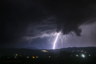
Lightning is a natural phenomenon formed by the occurrence of lightning bolts, which are electrostatic discharges through the atmosphere between two electrically charged regions, either both in the atmosphere or with one in the atmosphere and on the ground, temporarily neutralizing these in a near-instantaneous release of an average of one gigajoule of energy. This discharge may produce a wide range of electromagnetic radiation, from heat created by the rapid movement of electrons, to brilliant flashes of visible light in the form of black-body radiation. Lightning causes thunder, a sound from the shock wave which develops as gases in the vicinity of the discharge experience a sudden increase in pressure. Lightning occurs commonly during thunderstorms as well as other types of energetic weather systems, but volcanic lightning can also occur during volcanic eruptions. Lightning is an atmospheric electrical phenomenon and contributes to the global atmospheric electrical circuit.

Ball lightning is a rare and unexplained phenomenon described as luminescent, spherical objects that vary from pea-sized to several meters in diameter. Though usually associated with thunderstorms, the observed phenomenon is reported to last considerably longer than the split-second flash of a lightning bolt, and is a phenomenon distinct from St. Elmo's fire.

Functional magnetic resonance imaging or functional MRI (fMRI) measures brain activity by detecting changes associated with blood flow. This technique relies on the fact that cerebral blood flow and neuronal activation are coupled. When an area of the brain is in use, blood flow to that region also increases.

Medical imaging is the technique and process of imaging the interior of a body for clinical analysis and medical intervention, as well as visual representation of the function of some organs or tissues (physiology). Medical imaging seeks to reveal internal structures hidden by the skin and bones, as well as to diagnose and treat disease. Medical imaging also establishes a database of normal anatomy and physiology to make it possible to identify abnormalities. Although imaging of removed organs and tissues can be performed for medical reasons, such procedures are usually considered part of pathology instead of medical imaging.

Claustrophobia is the fear of confined spaces. It can be triggered by many situations or stimuli, including elevators, especially when crowded to capacity, windowless rooms, and hotel rooms with closed doors and sealed windows. Even bedrooms with a lock on the outside, small cars, and tight-necked clothing can induce a response in those with claustrophobia. It is typically classified as an anxiety disorder, which often results in panic attacks. The onset of claustrophobia has been attributed to many factors, including a reduction in the size of the amygdala, classical conditioning, or a genetic predisposition to fear small spaces.

Ferrofluid is a liquid that is attracted to the poles of a magnet. It is a colloidal liquid made of nanoscale ferromagnetic or ferrimagnetic particles suspended in a carrier fluid. Each magnetic particle is thoroughly coated with a surfactant to inhibit clumping. Large ferromagnetic particles can be ripped out of the homogeneous colloidal mixture, forming a separate clump of magnetic dust when exposed to strong magnetic fields. The magnetic attraction of tiny nanoparticles is weak enough that the surfactant's Van der Waals force is sufficient to prevent magnetic clumping or agglomeration. Ferrofluids usually do not retain magnetization in the absence of an externally applied field and thus are often classified as "superparamagnets" rather than ferromagnets.

A phosphene is the phenomenon of seeing light without light entering the eye. The word phosphene comes from the Greek words phos (light) and phainein. Phosphenes that are induced by movement or sound may be associated with optic neuritis.
The first neuroimaging technique ever is the so-called 'human circulation balance' invented by Angelo Mosso in the 1880s and able to non-invasively measure the redistribution of blood during emotional and intellectual activity. Then, in the early 1900s, a technique called pneumoencephalography was set. This process involved draining the cerebrospinal fluid from around the brain and replacing it with air, altering the relative density of the brain and its surroundings, to cause it to show up better on an x-ray, and it was considered to be incredibly unsafe for patients. A form of magnetic resonance imaging (MRI) and computed tomography (CT) were developed in the 1970s and 1980s. The new MRI and CT technologies were considerably less harmful and are explained in greater detail below. Next came SPECT and PET scans, which allowed scientists to map brain function because, unlike MRI and CT, these scans could create more than just static images of the brain's structure. Learning from MRI, PET and SPECT scanning, scientists were able to develop functional MRI (fMRI) with abilities that opened the door to direct observation of cognitive activities.

A lightning strike is a lightning event in which the electric discharge takes place between the atmosphere and the ground. Most originate in a cumulonimbus cloud and terminate on the ground, called cloud-to-ground (CG) lightning. A less common type of strike, ground-to-cloud (GC) lightning, is upward-propagating lightning initiated from a tall grounded object and reaching into the clouds. About 25% of all lightning events worldwide are strikes between the atmosphere and earth-bound objects. Most are intracloud (IC) lightning and cloud-to-cloud (CC), where discharges only occur high in the atmosphere. Lightning strikes the average commercial aircraft at least once a year, but modern engineering and design means this is rarely a problem. The movement of aircraft through clouds can even cause lightning strikes.

Neuroimaging is the use of quantitative (computational) techniques to study the structure and function of the central nervous system, developed as an objective way of scientifically studying the healthy human brain in a non-invasive manner. Increasingly it is also being used for quantitative studies of brain disease and psychiatric illness. Neuroimaging is a highly multidisciplinary research field and is not a medical specialty.
An earthquake light is a luminous aerial phenomenon that appears in the sky at or near areas of tectonic stress, seismic activity, or volcanic eruptions. There is no broad consensus as to the causes of the phenomenon involved. The phenomenon differs from disruptions to electrical grids – such as arcing power lines – which can produce bright flashes as a result of ground shaking or hazardous weather conditions.

A lightning detector is a device that detects lightning produced by thunderstorms. There are three primary types of detectors: ground-based systems using multiple antennas, mobile systems using a direction and a sense antenna in the same location, and space-based systems.
Preclinical imaging is the visualization of living animals for research purposes, such as drug development. Imaging modalities have long been crucial to the researcher in observing changes, either at the organ, tissue, cell, or molecular level, in animals responding to physiological or environmental changes. Imaging modalities that are non-invasive and in vivo have become especially important to study animal models longitudinally. Broadly speaking, these imaging systems can be categorized into primarily morphological/anatomical and primarily molecular imaging techniques. Techniques such as high-frequency micro-ultrasound, magnetic resonance imaging (MRI) and computed tomography (CT) are usually used for anatomical imaging, while optical imaging, positron emission tomography (PET), and single photon emission computed tomography (SPECT) are usually used for molecular visualizations.

Real-time magnetic resonance imaging (RT-MRI) refers to the continuous monitoring ("filming") of moving objects in real time. Because MRI is based on time-consuming scanning of k-space, real-time MRI was possible only with low image quality or low temporal resolution. Using an iterative reconstruction algorithm these limitations have recently been removed: a new method for real-time MRI achieves a temporal resolution of 20 to 30 milliseconds for images with an in-plane resolution of 1.5 to 2.0 mm. Real-time MRI promises to add important information about diseases of the joints and the heart. In many cases MRI examinations may become easier and more comfortable for patients.

Magnetic resonance imaging (MRI) is in general a safe technique, although injuries may occur as a result of failed safety procedures or human error. During the last 150 years, thousands of papers focusing on the effects or side effects of magnetic or radiofrequency fields have been published. They can be categorized as incidental and physiological. Contraindications to MRI include most cochlear implants and cardiac pacemakers, shrapnel and metallic foreign bodies in the eyes. The safety of MRI during the first trimester of pregnancy is uncertain, but it may be preferable to other options. Since MRI does not use any ionizing radiation, its use generally is favored in preference to CT when either modality could yield the same information.

An MRI sequence in magnetic resonance imaging (MRI) is a particular setting of pulse sequences and pulsed field gradients, resulting in a particular image appearance.

Portable magnetic resonance imaging (MRI) is referred to the imaging provided by an MRI scanner that has mobility and portability. It provides MR imaging to the patient in-time and on-site, for example, in Intensive care unit (ICU) where there is danger associated with moving the patient, in an ambulance, after a disaster rescue, or in a field hospital/medical tent.
References
- ↑ "ReviseMRI.com : Magnetophosphenes".
- ↑ Weintraub MI, Khoury A, Cole SP (July 2007). "Biologic effects of 3 Tesla (T) MR imaging comparing traditional 1.5 T and 0.6 T in 1023 consecutive outpatients". J Neuroimaging. 17 (3): 241–5. doi:10.1111/j.1552-6569.2007.00118.x. PMID 17608910. S2CID 44619313.
- ↑ "Technology Review: Blogs: arXiv blog: Magnetically Induced Hallucinations Explain Ball Lightning, Say Physicists". www.technologyreview.com. Archived from the original on 2010-05-13.