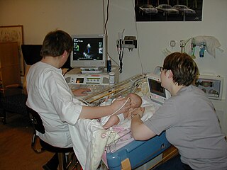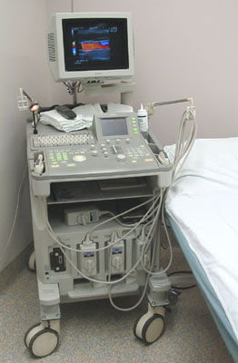
Medical ultrasound includes diagnostic techniques using ultrasound, as well as therapeutic applications of ultrasound. In diagnosis, it is used to create an image of internal body structures such as tendons, muscles, joints, blood vessels, and internal organs, to measure some characteristics or to generate an informative audible sound. The usage of ultrasound to produce visual images for medicine is called medical ultrasonography or simply sonography. The practice of examining pregnant women using ultrasound is called obstetric ultrasonography, and was an early development of clinical ultrasonography.

Radiology is the medical discipline that uses medical imaging to diagnose diseases and guide their treatment, within the bodies of humans and other animals. It began with radiography, but today it includes all imaging modalities, including those that use no electromagnetic radiation, as well as others that do, such as computed tomography (CT), fluoroscopy, and nuclear medicine including positron emission tomography (PET). Interventional radiology is the performance of usually minimally invasive medical procedures with the guidance of imaging technologies such as those mentioned above.
Medical physics deals with the application of the concepts and methods of physics to the prevention, diagnosis and treatment of human diseases with a specific goal of improving human health and well-being. Since 2008, medical physics has been included as a health profession according to International Standard Classification of Occupation of the International Labour Organization.

The human shoulder is made up of three bones: the clavicle (collarbone), the scapula, and the humerus as well as associated muscles, ligaments and tendons. The articulations between the bones of the shoulder make up the shoulder joints. The shoulder joint, also known as the glenohumeral joint, is the major joint of the shoulder, but can more broadly include the acromioclavicular joint. In human anatomy, the shoulder joint comprises the part of the body where the humerus attaches to the scapula, and the head sits in the glenoid cavity. The shoulder is the group of structures in the region of the joint.

Hospital for Special Surgery (HSS) is a hospital in New York City that specializes in orthopedic surgery and the treatment of rheumatologic conditions.

A sonographer is an allied healthcare professional who specializes in the use of ultrasonic imaging devices to produce diagnostic images, scans, videos or three-dimensional volumes of anatomy and diagnostic data. The requirements for clinical practice vary greatly by country. Sonography requires specialized education and skills to acquire, analyze and optimize information in the image. Due to the high levels of decisional latitude and diagnostic input, sonographers have a high degree of responsibility in the diagnostic process. Many countries require medical sonographers to have professional certification. Sonographers have core knowledge in ultrasound physics, cross-sectional anatomy, physiology, and pathology.

The Wayne State University School of Medicine (WSUSOM) is the medical school of Wayne State University, a public research university in Detroit, Michigan. It enrolls more than 1,500 students in undergraduate medical education, master's degree, Ph.D., and M.D.-Ph.D.. WSUSOM traces its roots through four predecessor institutions since its founding in 1868.
The American College of Radiology (ACR), founded in 1923, is a professional medical society representing nearly 40,000 diagnostic radiologists, radiation oncologists, interventional radiologists, nuclear medicine physicians and medical physicists.

Radiographers, also known as radiologic technologists, diagnostic radiographers and medical radiation technologists are healthcare professionals who specialize in the imaging of human anatomy for the diagnosis and treatment of pathology. Radiographers are infrequently, and almost always erroneously, known as x-ray technicians. In countries that use the title radiologic technologist they are often informally referred to as techs in the clinical environment; this phrase has emerged in popular culture such as television programmes. The term radiographer can also refer to a therapeutic radiographer, also known as a radiation therapist.

Abdominal ultrasonography is a form of medical ultrasonography to visualise abdominal anatomical structures. It uses transmission and reflection of ultrasound waves to visualise internal organs through the abdominal wall. For this reason, the procedure is also called a transabdominal ultrasound, in contrast to endoscopic ultrasound, the latter combining ultrasound with endoscopy through visualize internal structures from within hollow organs.
The American Registry for Diagnostic Medical Sonography (ARDMS), incorporated in June 1975, is an independent nonprofit organization that administers examinations and awards credentials in the areas of diagnostic medical sonography, diagnostic cardiac sonography, vascular technology, physicians’ vascular interpretation, musculoskeletal sonography and midwifery ultrasound. ARDMS has over 90,000 certified individuals in the U.S., Canada and throughout the world. ARDMS provides certifications, resources, and career information to healthcare practitioners and students practicing medical sonography.
The Advanced Diagnostic Ultrasound in Microgravity (ADUM) project is a U.S. government-funded study investigating strategies for applying diagnostic telemedicine to space. The Principal Investigator is Scott Dulchavsky, Chairman of Surgery at the Henry Ford Health System. This study was the first formal experiment to examine the use of ultrasound in microgravity encompassing musculoskeletal, heart, lung, abdominal, pelvic, dental, and orbital scans. Ultrasound is the only medical imaging device currently available on board the International Space Station. In addition, the lack of physician expertise on board the ISS makes diagnosis of medical conditions challenging. Ultrasound may have direct application for the evaluation and diagnosis of hundreds of medical conditions and is of interest for treating exploration crews. The telemedicine strategies investigated by this project has widespread application on Earth in emergency and rural care situations. Ultrasound images from space from a variety of body regions have been shown to be of diagnostic quality and non-expert operators were easily trained in ultrasound skills. This work has been expanded to include professional Football, Baseball, and Ice Hockey teams as well as the Winter and Summer Olympic Games in collaboration with investigators such as Marnix Van Holsbeeck. Dr. Dulchavsky has also led an innovative pilot study to expand comprehensive ultrasound education to basic science medical students at the Wayne State University School of Medicine. This trial has been shown to be a success with over 82 percent of students agreeing or strongly agreeing that their educational experience with the lightweight ultrasound technology education was positive. This technology is now being taught to medical students in their clinical clerkships.
Thomas M. Kolb, M.D., is an American radiologist specializing in the detection and diagnosis of breast cancer in young, predominantly high-risk premenopausal women. He has served as an assistant clinical professor of Radiology at Columbia University College of Physicians and Surgeons from 1994–2010. Dr. Kolb is double board certified, having received his training in pediatrics at the Albert Einstein College of Medicine in Bronx, New York, and in diagnostic radiology at the Columbia-Presbyterian Medical Center in New York.

AECC University College is a specialist university in Bournemouth that offers undergraduate, postgraduate, and short courses in a range of health sciences disciplines.
Mario Maas is a Dutch professor of radiology, in particular musculoskeletal radiology at the University of Amsterdam and the Academic Medical Centre of Amsterdam.

Ferenc Andras Jolesz was a Hungarian-American physician and scientist best known for his research on image guided therapy, the process by which information derived from diagnostic imaging is used to improve the localization and targeting of diseased tissue to monitor and control treatment during surgical and interventional procedures. He pioneered the field of Magnetic Resonance Imaging-guided interventions and introduced of a variety of new medical procedures based on novel combinations of imaging and therapy delivery.

Since its establishment in 1958, NASA has conducted research on a range of topics. Because of its unique structure, work happens at various field centers and different research areas are concentrated in those centers. Depending on the technology, hardware and expertise needed, research may be conducted across a range of centers.
Terason, a division of Teratech Corporation, is a global medical ultrasound imaging company headquartered in Burlington, Massachusetts, USA. Terason was the first to patent color portable ultrasound and is a market leader in ultrasound-guided venous intervention. Terason produces portable ultrasound products and technologies and has provided ultrasound systems to clinicians, hospitals, outpatient centers, and OEM partners since 2000.
Michael Grace Kawooya is a Ugandan physician, academic, researcher and academic administrator, who serves as Director at the Ernest Cook Ultrasound Research and Education Institute (ECUREI). He is a Professor (emeritus) of Radiology at Makerere University College of Health Sciences.
The Faculty of Sport and Exercise Medicine (UK) (FSEM) is a not-for-profit professional organisation responsible for training, educating and representing over 500 doctors in the United Kingdom. These doctors practise in the speciality of sport and exercise medicine (SEM). The FSEM is housed in the Royal College of Surgeons Edinburgh, but is an intercollegiate faculty of the Royal Colleges of Physicians and RCSEd


