Related Research Articles
Cranial nerves are the nerves that emerge directly from the brain, of which there are conventionally considered twelve pairs. Cranial nerves relay information between the brain and parts of the body, primarily to and from regions of the head and neck, including the special senses of vision, taste, smell, and hearing.
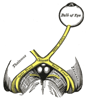
The optic nerve, also known as cranial nerve II, or simply as CN II, is a paired cranial nerve that transmits visual information from the retina to the brain. In humans, the optic nerve is derived from optic stalks during the seventh week of development and is composed of retinal ganglion cell axons and glial cells; it extends from the optic disc to the optic chiasma and continues as the optic tract to the lateral geniculate nucleus, pretectal nuclei, and superior colliculus.

The abducens nerve is the sixth cranial nerve (CNVI), in humans, that controls the movement of the lateral rectus muscle, responsible for outward gaze. It is a somatic efferent nerve.

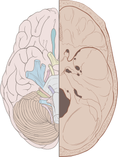
The brainstem is the posterior part of the brain, continuous with the spinal cord. In the human brain the brainstem is composed of the midbrain, the pons, and the medulla oblongata. The midbrain is continuous with the thalamus of the diencephalon through the tentorial notch, and sometimes the diencephalon is included in the brainstem.

The oculomotor nerve is the third cranial nerve. It enters the orbit via the superior orbital fissure and innervates extrinsic eye muscles that enable most movements of the eye and that raise the eyelid. The nerve also contains fibers that innervate the intrinsic eye muscles that enable pupillary constriction and accommodation. The oculomotor nerve is derived from the basal plate of the embryonic midbrain. Cranial nerves IV and VI also participate in control of eye movement.

The vestibulo-ocular reflex (VOR) is a reflex acting to stabilize gaze during head movement, with eye movement due to activation of the vestibular system. The reflex acts to stabilize images on the retinas of the eye during head movement, holding gaze is held steadily on a location, by producing eye movements in the direction opposite to head movement. For example, when the head moves to the right, the eyes move to the left, meaning the image a person sees stays the same even though the head has turned. Since slight head movement is present all the time, VOR is necessary for stabilizing vision: people with an impaired reflex find it difficult to read using print, because the eyes do not stabilise during small head tremors, and also because damage to reflex can cause nystagmus.

The pupillary light reflex (PLR) or photopupillary reflex is a reflex that controls the diameter of the pupil, in response to the intensity (luminance) of light that falls on the retinal ganglion cells of the retina in the back of the eye, thereby assisting in adaptation of vision to various levels of lightness/darkness. A greater intensity of light causes the pupil to constrict, whereas a lower intensity of light causes the pupil to dilate. Thus, the pupillary light reflex regulates the intensity of light entering the eye. Light shone into one eye will cause both pupils to constrict.
Blinking is a bodily function; it is a semi-autonomic rapid closing of the eyelid. A single blink is determined by the forceful closing of the eyelid or inactivation of the levator palpebrae superioris and the activation of the palpebral portion of the orbicularis oculi, not the full open and close. It is an essential function of the eye that helps spread tears across and remove irritants from the surface of the cornea and conjunctiva.
The optic tract is a part of the visual system in the brain. It is a continuation of the optic nerve that relays information from the optic chiasm to the ipsilateral lateral geniculate nucleus (LGN), pretectal nuclei, and superior colliculus.

The accommodation reflex is a reflex action of the eye, in response to focusing on a near object, then looking at a distant object, comprising coordinated changes in vergence, lens shape (accommodation) and pupil size. It is dependent on cranial nerve II, superior centers (interneuron) and cranial nerve III. The change in the shape of the lens is controlled by the ciliary muscles inside the eye. Changes in contraction of the ciliary muscles alter the focal distance of the eye, causing nearer or farther images to come into focus on the retina; this process is known as accommodation. The reflex, controlled by the parasympathetic nervous system, involves three responses: pupil constriction, lens accommodation, and convergence.
The pretectal area, or pretectum, is a midbrain structure composed of seven nuclei and comprises part of the subcortical visual system. Through reciprocal bilateral projections from the retina, it is involved primarily in mediating behavioral responses to acute changes in ambient light such as the pupillary light reflex, the optokinetic reflex, and temporary changes to the circadian rhythm. In addition to the pretectum's role in the visual system, the anterior pretectal nucleus has been found to mediate somatosensory and nociceptive information.

Exophthalmos is a bulging of the eye anteriorly out of the orbit. Exophthalmos can be either bilateral or unilateral. Complete or partial dislocation from the orbit is also possible from trauma or swelling of surrounding tissue resulting from trauma.
Posterior ischemic optic neuropathy (PION) is a medical condition characterized by damage to the retrobulbar portion of the optic nerve due to inadequate blood flow (ischemia) to the optic nerve. Despite the term posterior, this form of damage to the eye's optic nerve due to poor blood flow also includes cases where the cause of inadequate blood flow to the nerve is anterior, as the condition describes a particular mechanism of visual loss as much as the location of damage in the optic nerve. In contrast, anterior ischemic optic neuropathy (AION) is distinguished from PION by the fact that AION occurs spontaneously and on one side in affected individuals with predisposing anatomic or cardiovascular risk factors.
The corneal reflex, also known as the blink reflex, is an involuntary blinking of the eyelids elicited by stimulation of the cornea, though could result from any peripheral stimulus. Stimulation should elicit both a direct and consensual response. The reflex occurs at a rapid rate of 0.1 seconds. The purpose of this reflex is to protect the eyes from foreign bodies and bright lights. The blink reflex also occurs when sounds greater than 40–60 dB are made.

The nasociliary nerve is a branch of the ophthalmic nerve, itself a branch of the trigeminal nerve. It is intermediate in size between the other two branches of the ophthalmic nerve, the frontal nerve and lacrimal nerve.
The glabellar reflex, also known as the "glabellar tap sign", is a primitive reflex elicited by repetitive tapping on the forehead. Subjects blink in response to the first several taps. If the blinking persists, this is known as Myerson's sign, and is abnormal and a sign of frontal release; it is often seen in people who have Parkinson's disease.
Relative afferent pupillary defect (RAPD) is a medical sign observed during the swinging-flashlight test whereupon the patient's pupils dilate when a bright light is swung from the unaffected eye to the affected eye. The affected eye still senses the light and produces pupillary sphincter constriction to some degree, albeit reduced.
The optical pathway collects, transforms and creates an idea of the phenomena related to the presence and manifestations of light energy. Most of the impressions from the outside world are obtained through the senses of sight. The retina, which is the receiving part of the sense pathway of vision, as well as the optic nerve during development, arise from parts of the central nervous system. The sense of sight is well developed in terrestrial vertebrates with binocular vision. Primates are already developing the ability of three-dimensional vision, and humans have seen the most perfect vision, which is especially related to the high degree of development of the cerebral cortex, especially the associative optical cortex.

The visual pathway consists of structures that carry visual information from the retina to the brain. Lesions in that pathway cause a variety of visual field defects. In the visual system of human eye, the visual information processed by retinal photoreceptor cells travel in the following way:
Retina→Optic nerve→Optic chiasm →Optic tract→Lateral geniculate nucleus→Optic radiation→Primary and secondary visual cortices.
References
- 1 2 3 4 Francis Heed Adler (1953). Physiology of the eye: clinical application (2nd ed.). Mosby. pp. 23.
- 1 2 Douglas H. Slatter (2001). Fundamentals of veterinary ophthalmology (3rd ed.). Elsevier Health Sciences. p. 18. ISBN 9780721627052.
- 1 2 3 Michael Edward Peterson & Patricia A. Talcott (2006). Small animal toxicology (2nd ed.). Elsevier Health Sciences. p. 101. ISBN 9780721606392.
- ↑ O. M. Radostits; J. H. Arundel & Clive C. Gay (2000). O. M. Radostits (ed.). Veterinary medicine: a textbook of the diseases of cattle, sheep, pigs, goats and horses (9th ed.). Elsevier Health Sciences. p. 512. ISBN 9780702026041.
| This biology article is a stub. You can help Wikipedia by expanding it. |