Related Research Articles

The cerebral cortex, also known as the cerebral mantle, is the outer layer of neural tissue of the cerebrum of the brain in humans and other mammals. It is separated into two cortices, by the longitudinal fissure that divides the cerebrum into the left and right cerebral hemispheres. The two hemispheres are joined beneath the cortex by the corpus callosum. The cerebral cortex is the largest site of neural integration in the central nervous system. It plays a key role in attention, perception, awareness, thought, memory, language, and consciousness.

The limbic system, also known as the paleomammalian cortex, is a set of brain structures located on both sides of the thalamus, immediately beneath the medial temporal lobe of the cerebrum primarily in the midbrain.

A Brodmann area is a region of the cerebral cortex, in the human or other primate brain, defined by its cytoarchitecture, or histological structure and organization of cells.
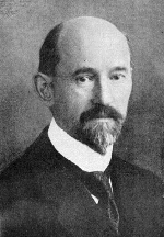
Korbinian Brodmann was a German neurologist who became famous for his definition of the cerebral cortex into 52 distinct regions from their cytoarchitectonic (histological) characteristics, known as Brodmann areas.
Proisocortex or pro-isocortex is one of two subtypes of cortical areas in the areas belonging to the neocortex. The other subtype is termed the true isocortex. Proisocortical areas are transitional areas placed between areas of true isocortex and areas of periallocortex. The histological structure of proisocortex is also transitional between true isocortex and either peripaleocortex or periarchicortex, depending on with which subtype of periallocortex the given proisocortical area borders.

The neocortex, also called the neopallium and isocortex, is the part of the mammalian brain involved in higher-order brain functions such as sensory perception, cognition, generation of motor commands, spatial reasoning and language.

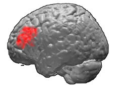
Brodmann area 10 is the anterior-most portion of the prefrontal cortex in the human brain. BA10 was originally defined broadly in terms of its cytoarchitectonic traits as they were observed in the brains of cadavers, but because modern functional imaging cannot precisely identify these boundaries, the terms anterior prefrontal cortex, rostral prefrontal cortex and frontopolar prefrontal cortex are used to refer to the area in the most anterior part of the frontal cortex that approximately covers BA10—simply to emphasize the fact that BA10 does not include all parts of the prefrontal cortex.
Brodmann area 46, or BA46, is part of the frontal cortex in the human brain. It is between BA10 and BA45.

In anatomy of animals, the archicortex or archipallium is the phylogenetically oldest region of the brain's pallium or cortex.
The allocortex is one of the two types of cerebral cortex, the other being the neocortex. It is characterized by having just three or four cell layers, in contrast with the six layers of the neocortex, and takes up a much smaller area than the neocortex. There are three subtypes of allocortex: the paleocortex, the archicortex, and the periallocortex – a transitional zone between the neocortex and the allocortex.

Cytoarchitecture, also known as cytoarchitectonics, is the study of the cellular composition of the central nervous system's tissues under the microscope. Cytoarchitectonics is one of the ways to parse the brain, by obtaining sections of the brain using a microtome and staining them with chemical agents which reveal where different neurons are located.

The posterior cingulate cortex (PCC) is the caudal part of the cingulate cortex, located posterior to the anterior cingulate cortex. This is the upper part of the "limbic lobe". The cingulate cortex is made up of an area around the midline of the brain. Surrounding areas include the retrosplenial cortex and the precuneus.

In anatomy of animals, the paleocortex, or paleopallium is a region within the telencephalon in the brain which is older in an evolutionary sense than the archicortex and the neocortex.
The paralimbic cortex is an area of three-layered cortex that includes the following regions: the piriform cortex, entorhinal cortex, the parahippocampal cortex on the medial surface of the temporal lobe, and the cingulate cortex just above the corpus callosum.

In neuroanatomy, pallium refers to the layers of grey and white matter that cover the upper surface of the cerebrum in vertebrates. The non-pallial part of the telencephalon builds the subpallium. In basal vertebrates the pallium is a relatively simple three-layered structure, encompassing 3-4 histogenetically distinct domains, plus the olfactory bulb. It used to be thought that pallium equals cortex and subpallium equals telencephalic nuclei, but it has turned out, according to comparative evidence provided by molecular markers, that the pallium develops both cortical structures and pallial nuclei, whereas the subpallium develops striatal, pallidal, diagonal-innominate and preoptic nuclei, plus the corticoid structure of the olfactory tuberculum. In mammals, the cortical part of the pallium registers a definite evolutionary step-up in complexity, forming the cerebral cortex, most of which consists of a progressively expanded six-layered portion isocortex, with simpler three-layered cortical regions allocortex at the margins. The allocortex subdivides into hippocampal allocortex, medially, and olfactory allocortex, laterally.
Agranular insula is a portion of the cerebral cortex defined on the basis of internal structure in the human, the macaque, the rat, and the mouse. Classified as allocortex (periallocortex), it is in primates distinguished from adjacent neocortex (proisocortex) by absence of the external granular layer (II) and of the internal granular layer (IV). It occupies the anterior part of the insula, the posterior portion of the orbital gyri and the medial part of the temporal pole. In rodents it is located on the ventrolateral surface of the cortex rostrally, between the piriform area ventrally and the gustatory area or the visceral area dorsally.
Granular insular cortex refers to a portion of the cerebral cortex defined on the basis of internal structure in the human and macaque, the rat, and the mouse. Classified as neocortex, it is in primates distinguished from adjacent allocortex (periallocortex) by the presence of granular layers – external granular layer (II) and internal granular layer (IV) – and by differentiation of the external pyramidal layer (III) into sublayers. In primates it occupies the posterior part of the insula. In rodents it is located on the lateral surface of the cortex rostrally, dorsal to the gustatory area or, more caudally, dorsal to the agranular insula.
Periallocortex is one of three subtypes of allocortex, the other two subtypes being paleocortex and archicortex. The periallocortex is formed at transition areas where any of the other two subtypes of allocortex borders with the neocortex.
Peripaleocortex is one of two subtypes of periallocortex, the other being periarchicortex. Peripaleocortex is formed at borders between isocortex (neocortex) and paleocortex. It shows slow histological transition from the three-layered structure characteristic of paleocortex to the typical six-layered structure characteristic of isocortex. The main peripaleocortex area is anterior insular cortex.
Periarchicortex is one of two subtypes of periallocortex, the other being peripaleocortex. It is formed at borders between archicortex and isocortex and shows slow histological transition from the four-layered structure typical for archicortex to the six-layered structure typical for isocortex.
References
- ↑ mediLexicon: Definition: 'Juxtallocortex'. http://www.medilexicon.com/medicaldictionary.php?t=46602
- ↑ Mesulam, 2000 In: M.M. Mesulam, Editor, Principles of Behavioral and Cognitive Neurology (2nd ed.), Oxford University Press, New York (2000)
- 1 2 "Mesocortex". BrainInfo. University of Washington. Retrieved 13 October 2017.
- ↑ G. Avanzini, Anne Beaumanoir, Laura Mira, eds. Limbic Seizures in Children. John Libbey Eurotext, 2001, p. 13
- ↑ Almut Schuez, Robert Miller. Cortical Areas: Unity and Diversity. CRC Press, 13 Jul 2003, pp. 236-237.
- ↑ Reep R. Relationship between prefrontal and limbic cortex: a comparative anatomical review. Brain Behav Evol. 1984;25(1):5-80.