Related Research Articles

The pudendal nerve is the main nerve of the perineum. It carries sensation from the external genitalia of both sexes and the skin around the anus and perineum, as well as the motor supply to various pelvic muscles, including the male or female external urethral sphincter and the external anal sphincter. If damaged, most commonly by childbirth, lesions may cause sensory loss or fecal incontinence. The nerve may be temporarily blocked as part of an anaesthetic procedure.

In human anatomy, the ulnar nerve is a nerve that runs near the ulna bone. The ulnar collateral ligament of elbow joint is in relation with the ulnar nerve. The nerve is the largest in the human body unprotected by muscle or bone, so injury is common. This nerve is directly connected to the little finger, and the adjacent half of the ring finger, innervating the palmar aspect of these fingers, including both front and back of the tips, perhaps as far back as the fingernail beds.

The ligamentum arteriosum, also known as the Ligament of Botallo or Harvey's ligament, is a small ligament attaching the aorta to the pulmonary artery. It serves no function in adults but is the remnant of the ductus arteriosus formed within three weeks after birth.
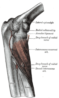
In human anatomy, the supinator is a broad muscle in the posterior compartment of the forearm, curved around the upper third of the radius. Its function is to supinate the forearm.

The superior gluteal nerve is a nerve that originates in the pelvis. It supplies the gluteus medius muscle, the gluteus minimus muscle, the tensor fasciae latae muscle, and the piriformis muscle.

The obturator nerve in human anatomy arises from the ventral divisions of the second, third, and fourth lumbar nerves in the lumbar plexus; the branch from the third is the largest, while that from the second is often very small.
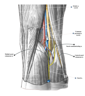
The sural nerve(L4-S1) is generally considered a pure cutaneous nerve of the posterolateral leg to the lateral ankle. The sural nerve originates from a combination of either the sural communicating branch and medial sural cutaneous nerve, or the lateral sural cutaneous nerve. This group of nerves is termed the sural nerve complex. There are eight documented variations of the sural nerve complex. Once formed the sural nerve takes its course midline posterior to posterolateral around the lateral malleolus. The sural nerve terminates as the lateral dorsal cutaneous nerve.

The supratrochlear nerve is a branch of the frontal nerve, itself a branch of the ophthalmic nerve (CN V1) from the trigeminal nerve (CN V). It provides sensory innervation to the skin of the forehead and the upper eyelid.
Pancreatic polypeptide (PP) is a polypeptide secreted by PP cells in the endocrine pancreas. It regulates pancreatic secretion activities, and also impacts liver glycogen storage and gastrointestinal secretion. Its secretion may be impacted by certain endocrine tumours.
The perineal nerve is a nerve of the pelvis. It arises from the pudendal nerve in the pudendal canal. It gives superficial branches to the skin, and a deep branch to muscles. It supplies the skin and muscles of the perineum. Its latency is tested with electrodes.

The lateral pectoral nerve arises from the lateral cord of the brachial plexus, and through it from the C5-7.

The angular vein is a vein of the face. It is the upper part of the facial vein, above its junction with the superior labial vein. It is formed by the junction of the supratrochlear vein and supraorbital vein, and joins with the superior labial vein. It drains the medial canthus, and parts of the nose and the upper lip. It can be a route of spread of infection from the danger triangle of the face to the cavernous sinus.

The tympanic nerve is a branch of the glossopharyngeal nerve found near the ear. It gives sensation to the middle ear, the Eustachian tube, the parotid gland, and mastoid air cells. It gives parasympathetic to supply to the parotid gland via the otic ganglion and the auriculotemporal nerve.

The posterior auricular nerve is a nerve of the head. It is a branch of the facial nerve. It communicates with branches from the vagus nerve, the great auricular nerve, and the lesser occipital nerve. Its auricular branch supplies the posterior auricular muscle, the intrinsic muscles of the auricle, and gives sensation to the auricle. Its occipital branch supplies the occipitalis muscle.

A vagotomy is a surgical procedure that involves removing part of the vagus nerve.
The zygomatic branches of the facial nerve (malar branches) are nerves of the face. They run across the zygomatic bone to the lateral angle of the orbit. Here, they supply the orbicularis oculi muscle, and join with filaments from the lacrimal nerve and the zygomaticofacial branch of the maxillary nerve (CN V2).
The cervical branch of the facial nerve is a nerve in the neck. It is a branch of the facial nerve (VII). It supplies the platysma muscle, among other functions.

The perineurium is a protective sheath that surrounds a nerve fascicle. This bundles together axons targeting the same anatomical location. The perineurium is composed from fibroblasts.

The respiratory center is located in the medulla oblongata and pons, in the brainstem. The respiratory center is made up of three major respiratory groups of neurons, two in the medulla and one in the pons. In the medulla they are the dorsal respiratory group, and the ventral respiratory group. In the pons, the pontine respiratory group includes two areas known as the pneumotaxic centre and the apneustic centre.
Post-vagotomy diarrhea is a form of diarrhea which occurs in 10% of people after a truncal vagotomy, which can range from severe to debilitating in approximately 2% to 4% of patients. However, the occurrence of post-vagotomy diarrhea is significantly reduced after proximal selective vagotomy, specifically when celiac and hepatic branches of the vagus are retained.
References
- ↑ Ellis, Harold; Mahadevan, Vishy (2010). "Peritoneal cavity". Clinical Anatomy: Applied Anatomy for Students and Junior Doctors (12 ed.). John Wiley and Sons. p. 81. ISBN 978-1-4051-8617-9.
- Schwartz's Principles of Surgery, Ninth Edition Book ISBN 978-0-07-1547703
| | This neuroanatomy article is a stub. You can help Wikipedia by expanding it. |