Related Research Articles

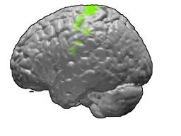
The motor cortex is the region of the cerebral cortex involved in the planning, control, and execution of voluntary movements. The motor cortex is an area of the frontal lobe located in the posterior precentral gyrus immediately anterior to the central sulcus.
Oligodendrocyte progenitor cells (OPCs), also known as oligodendrocyte precursor cells, NG2-glia, O2A cells, or polydendrocytes, are a subtype of glia in the central nervous system named for their essential role as precursors to oligodendrocytes. They are typically identified by coexpression of PDGFRA and NG2.

The premotor cortex is an area of the motor cortex lying within the frontal lobe of the brain just anterior to the primary motor cortex. It occupies part of Brodmann's area 6. It has been studied mainly in primates, including monkeys and humans. The functions of the premotor cortex are diverse and not fully understood. It projects directly to the spinal cord and therefore may play a role in the direct control of behavior, with a relative emphasis on the trunk muscles of the body. It may also play a role in planning movement, in the spatial guidance of movement, in the sensory guidance of movement, in understanding the actions of others, and in using abstract rules to perform specific tasks. Different subregions of the premotor cortex have different properties and presumably emphasize different functions. Nerve signals generated in the premotor cortex cause much more complex patterns of movement than the discrete patterns generated in the primary motor cortex.

The supplementary motor area (SMA) is a part of the motor cortex of primates that contributes to the control of movement. It is located on the midline surface of the hemisphere just in front of the primary motor cortex leg representation. In monkeys the SMA contains a rough map of the body. In humans the body map is not apparent. Neurons in the SMA project directly to the spinal cord and may play a role in the direct control of movement. Possible functions attributed to the SMA include the postural stabilization of the body, the coordination of both sides of the body such as during bimanual action, the control of movements that are internally generated rather than triggered by sensory events, and the control of sequences of movements. All of these proposed functions remain hypotheses. The precise role or roles of the SMA is not yet known.
Post-chemotherapy cognitive impairment (PCCI) describes the cognitive impairment that can result from chemotherapy treatment. Approximately 20 to 30% of people who undergo chemotherapy experience some level of post-chemotherapy cognitive impairment. The phenomenon first came to light because of the large number of breast cancer survivors who complained of changes in memory, fluency, and other cognitive abilities that impeded their ability to function as they had pre-chemotherapy.

The posterior parietal cortex plays an important role in planned movements, spatial reasoning, and attention.
Epileptogenesis is the gradual process by which a typical brain develops epilepsy. Epilepsy is a chronic condition in which seizures occur. These changes to the brain occasionally cause neurons to fire in an abnormal, hypersynchronous manner, known as a seizure.

A HEAT repeat is a protein tandem repeat structural motif composed of two alpha helices linked by a short loop. HEAT repeats can form alpha solenoids, a type of solenoid protein domain found in a number of cytoplasmic proteins. The name "HEAT" is an acronym for four proteins in which this repeat structure is found: Huntingtin, elongation factor 3 (EF3), protein phosphatase 2A (PP2A), and the yeast kinase TOR1. HEAT repeats form extended superhelical structures which are often involved in intracellular transport; they are structurally related to armadillo repeats. The nuclear transport protein importin beta contains 19 HEAT repeats.

The primary motor cortex is a brain region that in humans is located in the dorsal portion of the frontal lobe. It is the primary region of the motor system and works in association with other motor areas including premotor cortex, the supplementary motor area, posterior parietal cortex, and several subcortical brain regions, to plan and execute voluntary movements. Primary motor cortex is defined anatomically as the region of cortex that contains large neurons known as Betz cells, which, along with other cortical neurons, send long axons down the spinal cord to synapse onto the interneuron circuitry of the spinal cord and also directly onto the alpha motor neurons in the spinal cord which connect to the muscles.
Stuart C. Sealfon, M.D., is an American neurologist who studies the mechanisms of both the therapeutic and adverse effects of drugs. He was an early adopter of the use of massively parallel qPCR and fluorescent in situ hybridization to characterize cell response state and his research accomplishments have included the identification of the primary structure of the gonadotropin-releasing hormone receptor, finding new signaling pathways activated by drugs for Parkinson's disease, elucidating the mechanism of action of hallucinogens and finding a new brain receptor complex implicated in schizophrenia as a novel target for antipsychotics.
Noradrenergic cell group A1 is a group of cells in the vicinity of the lateral reticular nucleus of the medullary reticular formation that label for norepinephrine in primates and rodents. They are found in the ventrolateral medulla in conjunction with the adrenergic cell group C1.
Noradrenergic cell group A4 is a group of cells exhibiting noradrenergic fluorescence that, in the rat, are located in the Tegmen ventriculi quarti ventral to the cerebellar nuclei, and in the macaque, are found at the edge of the lateral recess of the fourth ventricle caudally, extending to beneath the floor of the ventricle where they merge with the noradrenergic group A6, the locus ceruleus.
Noradrenergic cell group A6 is a group of cells fluorescent for noradrenaline that are identical with the locus ceruleus, as identified by Nissl stain.
Noradrenergic cell group A6sc is a group of cells fluorescent for norepinephrine that are scattered in the nucleus subceruleus of the macaque.,
Noradrenergic cell group Acg is a group of cells fluorescent for norepinephrine that are located in the central gray of the midbrain at the level of the trochlear nucleus in the squirrel monkey (Saimiri) and to a lesser degree in the macaque.
Dopaminergic cell groups, DA cell groups, or dopaminergic nuclei are collections of neurons in the central nervous system that synthesize the neurotransmitter dopamine. In the 1960s, dopaminergic neurons or dopamine neurons were first identified and named by Annica Dahlström and Kjell Fuxe, who used histochemical fluorescence. The subsequent discovery of genes encoding enzymes that synthesize dopamine, and transporters that incorporate dopamine into synaptic vesicles or reclaim it after synaptic release, enabled scientists to identify dopaminergic neurons by labeling gene or protein expression that is specific to these neurons.
Serotonergic cell groups refer to collections of neurons in the central nervous system that have been demonstrated by histochemical fluorescence to contain the neurotransmitter serotonin (5-hydroxytryptamine). Since they are for the most part localized to classical brainstem nuclei, particularly the raphe nuclei, they are more often referred to by the names of those nuclei than by the B1-9 nomenclature. These cells appear to be common across most mammals and have two main regions in which they develop; one forms in the mesencephlon and the rostral pons and the other in the medulla oblongata and the caudal pons.

Pain is an aversive sensation and feeling associated with actual, or potential, tissue damage. It is widely accepted by a broad spectrum of scientists and philosophers that non-human animals can perceive pain, including pain in amphibians.
Santosh Gajanan Honavar is an Indian ophthalmologist and is currently the editor of the Indian Journal of Ophthalmology and Indian Journal of Ophthalmology - Case Reports, the official journals of the All India Ophthalmological Society; Director, Medical Services ; Director, Department of Ocular Oncology and Oculoplasty at Centre for Sight, Hyderabad; and Director, National Retinoblastoma Foundation.
David Rowitch, FMedSci, FRS is an American physician-scientist known for his contributions to developmental glial biology and treatment of white matter diseases. He heads the Department of Paediatrics at the University of Cambridge and is an adjunct professor of pediatrics at the University of California San Francisco (UCSF).
References
- ↑ Felten DL; Sladek JR Jr. (1983). "Monoamine distribution in primate brain V. Monoaminergic nuclei: anatomy, pathways and local organization". Brain Research Bulletin. 10 (2): 171–284. doi:10.1016/0361-9230(83)90045-x. PMID 6839182. S2CID 13176814.
{{cite journal}}: CS1 maint: multiple names: authors list (link) - ↑ Dahlstrom A; Fuxe K (1964). "Evidence for the existence of monoamine-containing neurons in the central nervous system". Acta Physiologica Scandinavica. 62: 1–55. PMID 14229500.
{{cite journal}}: CS1 maint: multiple names: authors list (link)