Related Research Articles

The central nervous system (CNS) is the part of the nervous system consisting primarily of the brain and spinal cord. The CNS is so named because the brain integrates the received information and coordinates and influences the activity of all parts of the bodies of bilaterally symmetric and triploblastic animals—that is, all multicellular animals except sponges and diploblasts. It is a structure composed of nervous tissue positioned along the rostral to caudal axis of the body and may have an enlarged section at the rostral end which is a brain. Only arthropods, cephalopods and vertebrates have a true brain.

A neurotransmitter is a signaling molecule secreted by a neuron to affect another cell across a synapse. The cell receiving the signal, any main body part or target cell, may be another neuron, but could also be a gland or muscle cell.

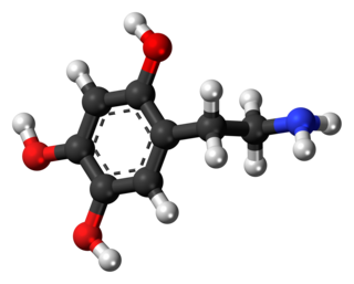
A catecholamine is a monoamine neurotransmitter, an organic compound that has a catechol and a side-chain amine.

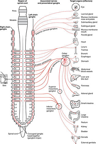
The autonomic nervous system (ANS), formerly referred to as the vegetative nervous system, is a division of the peripheral nervous system that supplies internal organs, smooth muscle and glands. The autonomic nervous system is a control system that acts largely unconsciously and regulates bodily functions, such as the heart rate, its force of contraction, digestion, respiratory rate, pupillary response, urination, and sexual arousal. This system is the primary mechanism in control of the fight-or-flight response.

The sympathetic nervous system (SNS) is one of the three divisions of the autonomic nervous system, the others being the parasympathetic nervous system and the enteric nervous system. The enteric nervous system is sometimes considered part of the autonomic nervous system, and sometimes considered an independent system.

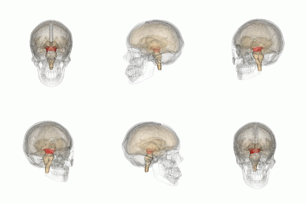
The brainstem is the posterior stalk-like part of the brain that connects the cerebrum with the spinal cord. In the human brain the brainstem is composed of the midbrain, the pons, and the medulla oblongata. The midbrain is continuous with the thalamus of the diencephalon through the tentorial notch, and sometimes the diencephalon is included in the brainstem.

In anatomy, the extrapyramidal system is a part of the motor system network causing involuntary actions. The system is called extrapyramidal to distinguish it from the tracts of the motor cortex that reach their targets by traveling through the pyramids of the medulla. The pyramidal tracts may directly innervate motor neurons of the spinal cord or brainstem, whereas the extrapyramidal system centers on the modulation and regulation of anterior (ventral) horn cells.

The raphe nuclei are a moderate-size cluster of nuclei found in the brain stem. They have 5-HT1 receptors which are coupled with Gi/Go-protein-inhibiting adenyl cyclase. They function as autoreceptors in the brain and decrease the release of serotonin. The anxiolytic drug Buspirone acts as partial agonist against these receptors. Selective serotonin reuptake inhibitor (SSRI) antidepressants are believed to act in these nuclei, as well as at their targets.

The locus coeruleus (LC), also spelled locus caeruleus or locus ceruleus, is a nucleus in the pons of the brainstem involved with physiological responses to stress and panic. It is a part of the reticular activating system.

The reticular formation is a set of interconnected nuclei that are located throughout the brainstem. It is not anatomically well defined, because it includes neurons located in different parts of the brain. The neurons of the reticular formation make up a complex set of networks in the core of the brainstem that extend from the upper part of the midbrain to the lower part of the medulla oblongata. The reticular formation includes ascending pathways to the cortex in the ascending reticular activating system (ARAS) and descending pathways to the spinal cord via the reticulospinal tracts.

The flocculus is a small lobe of the cerebellum at the posterior border of the middle cerebellar peduncle anterior to the biventer lobule. Like other parts of the cerebellum, the flocculus is involved in motor control. It is an essential part of the vestibulo-ocular reflex, and aids in the learning of basic motor skills in the brain.

Norepinephrine (NE), also called noradrenaline (NA) or noradrenalin, is an organic chemical in the catecholamine family that functions in the brain and body as both a hormone and neurotransmitter. The name "noradrenaline" is more commonly used in the United Kingdom, whereas "norepinephrine" is usually preferred in the United States. "Norepinephrine" is also the international nonproprietary name given to the drug. Regardless of which name is used for the substance itself, parts of the body that produce or are affected by it are referred to as noradrenergic.

Oxidopamine, also known as 6-hydroxydopamine (6-OHDA) or 2,4,5-trihydroxyphenethylamine, is a neurotoxic synthetic organic compound used by researchers to selectively destroy dopaminergic and noradrenergic neurons in the brain.
Noradrenergic cell group A7 is a group of cells fluorescent for norepinephrine that is located in the pontine reticular formation ventral to the superior cerebellar peduncle of the pons in rodents and in primates.
Catecholaminergic cell groups refers to collections of neurons in the central nervous system that have been demonstrated by histochemical fluorescence to contain one of the neurotransmitters dopamine or norepinephrine. Thus, it represents the combination of dopaminergic cell groups and noradrenergic cell groups. Some authors include in this category 'putative' adrenergic cell groups, collections of neurons that stain for PNMT, the enzyme that converts norepinephrine to epinephrine (adrenalin).
Dopaminergic cell groups, DA cell groups, or dopaminergic nuclei are collections of neurons in the central nervous system that synthesize the neurotransmitter dopamine. In the 1960s, dopaminergic neurons or dopamine neurons were first identified and named by Annica Dahlström and Kjell Fuxe, who used histochemical fluorescence. The subsequent discovery of genes encoding enzymes that synthesize dopamine, and transporters that incorporate dopamine into synaptic vesicles or reclaim it after synaptic release, enabled scientists to identify dopaminergic neurons by labeling gene or protein expression that is specific to these neurons.
Noradrenergic cell groups refers to collections of neurons in the central nervous system that have been demonstrated by histochemical fluorescence to contain the neurotransmitter norepinephrine (noradrenalin). They are named
Serotonergic cell groups refer to collections of neurons in the central nervous system that have been demonstrated by histochemical fluorescence to contain the neurotransmitter serotonin (5-hydroxytryptamine). Since they are for the most part localized to classical brainstem nuclei, particularly the raphe nuclei, they are more often referred to by the names of those nuclei than by the B1-9 nomenclature. These cells appear to be common across most mammals and have two main regions in which they develop; one forms in the mesencephlon and the rostral pons and the other in the medulla oblongata and the caudal pons.
The parabrachial nuclei, also known as the parabrachial complex, are a group of nuclei in the dorsolateral pons that surrounds the superior cerebellar peduncle as it enters the brainstem from the cerebellum. They are named from the Latin term for the superior cerebellar peduncle, the brachium conjunctivum. In the human brain, the expansion of the superior cerebellar peduncle expands the parabrachial nuclei, which form a thin strip of grey matter over most of the peduncle. The parabrachial nuclei are typically divided along the lines suggested by Baxter and Olszewski in humans, into a medial parabrachial nucleus and lateral parabrachial nucleus. These have in turn been subdivided into a dozen subnuclei: the superior, dorsal, ventral, internal, external and extreme lateral subnuclei; the lateral crescent and subparabrachial nucleus along the ventrolateral margin of the lateral parabrachial complex; and the medial and external medial subnuclei

The mesencephalic locomotor region (MLR) is a functionally defined area of the midbrain that is associated with the initiation and control of locomotor movements in vertebrate species.
References
- ↑ Felten DL, Sladek JR Jr (1983). "Monoamine distribution in primate brain V. Monoaminergic nuclei: anatomy, pathways and local organization". Brain Research Bulletin. 10 (2): 171–284. doi:10.1016/0361-9230(83)90045-x. PMID 6839182. S2CID 13176814.
- ↑ Dahlstrom A, Fuxe K (1964). "Evidence for the existence of monoamine-containing neurons in the central nervous system". Acta Physiologica Scandinavica. 62: 1–55. PMID 14229500.
- ↑ Kitahama K, Nagatsu I, Pearson J (1994). Catecholamine systems in mammalian midbrain and hindbrain: theme and variations. Chapter 8 in Phylogeny and Development of Catecholamine Systems in the CNS of Vertebrates, Smeets WJAJ and Reiner A (eds). Cambridge, England: University Press.
- ↑ Byrum, C.E. and Guyenet, P.G., 1987. Afferent and efferent connections of the A5 noradrenergic cell group in the rat. Journal of Comparative Neurology, 261(4), pp.529-542
- ↑ Woodruff, Michael L., Ronald H. Baisden, Dennis L. Whittington, and Joseph E. Kelly. "Inputs to the pontine A5 noradrenergic cell group: a horseradish peroxidase study." Experimental neurology 94, no. 3 (1986): 782-787
- ↑ Blessing, W. W., A. Ko Goodchild, R. A. L. Dampney, and J. Po Chalmers. "Cell groups in the lower brain stem of the rabbit projecting to the spinal cord, with special reference to catecholamine-containing neurons." Brain research 221, no. 1 (1981): 35-55
- ↑ Andrade, Rodrigo, and George K. Aghajanian. "Single cell activity in the noradrenergic A-5 region: responses to drugs and peripheral manipulations of blood pressure." Brain Research 242, no. 1 (1982) 125-136.
- ↑ Drye, Randall G., Ronald H. Baisden, Dennis L. Whittington, and Michael L. Woodruff. "The effects of stimulation of the A5 region on blood pressure and heart rate in rabbits." Brain research bulletin 24, no. 1 (1990) 33-39.
- ↑ Guyenet, P. G., Koshiya, N., Huangfu, G., Verberne, A. J. and Riley, T. A. "Central respiratory control of A5 and A6 pontine noradrenergic neurons." American Journal of Physiology. Regulatory, Integrative and Comparative Physiology 264, no. 6 (1993): R1035-R1044
- ↑ Taxini, Camila L., Thiago S. Moreira, Ana C. Takakura, Kênia C. Bícego, Luciane H. Gargaglioni, and Daniel B. Zoccal. "Role of A5 noradrenergic neurons in the chemoreflex control of respiratory and sympathetic activities in unanesthetized conditions." Neuroscience 354 (2017): 146-157.
- ↑ Woodruff, Michael L., Ronald H. Baisden, and Dennis L. Whittington. "Effects of electrical stimulation of the pontine A5 cell group on blood pressure and heart rate in the rabbit." Brain research 379, no. 1 (1986): 10-23.
- ↑ Pertovaara, Antti. "Noradrenergic pain modulation." Progress in neurobiology 80, no. 2 (2006): 53-83.