Oblique muscle of abdomen may refer to:
| This disambiguation page lists articles associated with the title Oblique muscle of abdomen. If an internal link led you here, you may wish to change the link to point directly to the intended article. |
Oblique muscle of abdomen may refer to:
| This disambiguation page lists articles associated with the title Oblique muscle of abdomen. If an internal link led you here, you may wish to change the link to point directly to the intended article. |
Oblique may refer to:

The rectus abdominis muscle, also known as the "abdominal muscle" or "abs", is a paired muscle running vertically on each side of the anterior wall of the human abdomen, as well as that of some other mammals. There are two parallel muscles, separated by a midline band of connective tissue called the linea alba. It extends from the pubic symphysis, pubic crest and pubic tubercle inferiorly, to the xiphoid process and costal cartilages of ribs V to VII superiorly. The proximal attachments are the pubic crest and the pubic symphysis. It attaches distally at the costal cartilages of ribs 5-7 and the xiphoid process of the sternum.

The transverse abdominal muscle (TVA), also known as the transverse abdominis, transversalis muscle and transversus abdominis muscle, is a muscle layer of the anterior and lateral abdominal wall which is deep to the internal oblique muscle. It is thought by most fitness instructors to be a significant component of the core.
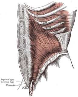
The abdominal internal oblique muscle, also internal oblique muscle or interior oblique, is an abdominal muscle in the abdominal wall that lies below the external oblique muscle and just above the transverse abdominal muscle.
The abdominal external oblique muscle is the largest and outermost of the three flat abdominal muscles of the lateral anterior abdomen.
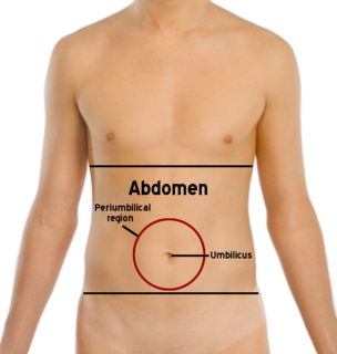
The abdomen is the part of the body between the thorax (chest) and pelvis, in humans and in other vertebrates. The abdomen is the front part of the abdominal segment of the trunk. The region occupied by the abdomen is called the abdominal cavity. In arthropods it is the posterior tagma of the body; it follows the thorax or cephalothorax.

In anatomy, the abdominal wall represents the boundaries of the abdominal cavity. The abdominal wall is split into the anterolateral and posterior walls.

The lumbar plexus is a web of nerves in the lumbar region of the body which forms part of the larger lumbosacral plexus. It is formed by the divisions of the first four lumbar nerves (L1-L4) and from contributions of the subcostal nerve (T12), which is the last thoracic nerve. Additionally, the ventral rami of the fourth lumbar nerve pass communicating branches, the lumbosacral trunk, to the sacral plexus. The nerves of the lumbar plexus pass in front of the hip joint and mainly support the anterior part of the thigh.
The iliohypogastric nerve is a nerve that originates from the lumbar plexus that supplies sensation to skin over the lateral gluteal region and motor to the internal oblique and transverse abdominal muscles.
The ilioinguinal nerve is a branch of the first lumbar nerve (L1). It separates from the first lumbar nerve along with the larger iliohypogastric nerve. It emerges from the lateral border of the psoas major just inferior to the iliohypogastric, and passes obliquely across the quadratus lumborum and iliacus. The ilioinguinal nerve then perforates the transversus abdominis near the anterior part of the iliac crest, and communicates with the iliohypogastric nerve between the transversus and the internal oblique muscle.

The conjoint tendon is a structure formed from the lower part of the common aponeurosis of the internal oblique muscle and the transversus abdominis as it inserts into the crest of the pubis and pectineal line immediately behind the superficial inguinal ring. It is usually conjoint with the tendon of the internal oblique muscle, but they may be separate as well. It forms the medial part of the posterior wall of the inguinal canal.
Abdominal exercises are those that affect the abdominal muscles.

The crest of the ilium is the superior border of the wing of ilium and the superiolateral margin of the greater pelvis.
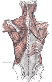
The lumbar triangle can refer to either the inferior lumbar (Petit) triangle, which lies superficially, or the superior lumbar (Grynfeltt) triangle, which is deep and superior to the inferior triangle. Of the two, the superior triangle is the more consistently found in cadavers, and is more commonly the site of herniation; however, the inferior lumbar triangle is often simply called the lumbar triangle, perhaps owing to its more superficial location and ease in demonstration.
The arcuate line of rectus sheath, linea semicircularis, arcuate line, or Douglas' line is a horizontal line that demarcates the lower limit of the posterior layer of the rectus sheath. It is also where the inferior epigastric vessels perforate the rectus abdominis.
The rectus sheath, also called the rectus fascia, is formed by the aponeuroses of the transverse abdominal and the internal and external oblique muscles. It contains the rectus abdominis and pyramidalis muscles.

The muscles of respiration are those muscles that contribute to inhalation and exhalation, by aiding in the expansion and contraction of the thoracic cavity. The diaphragm and, to a lesser extent, the intercostal muscles drive respiration during quiet breathing. Additional 'accessory muscles of respiration' are typically only used under conditions of high metabolic demand or respiratory dysfunction. However, in instances where these accessory muscles become stiff and hard, expansion of the rib cage can be restricted. Maintenance of the elasticity of these muscles is crucial to the health of the respiratory system and to maximize its functional capabilities.

The iliac branch of the iliolumbar artery descends to supply the iliacus muscle; some offsets, running between the muscle and the bone, anastomose with the iliac branches of the obturator artery; one of these enters an oblique canal to supply the bone, while others run along the crest of the ilium, distributing branches to the gluteal and abdominal muscles, and anastomosing in their course with the superior gluteal artery, iliac circumflex artery, and the lateral circumflex femoral artery. This anastamosis occurs around the anterior superior iliac spine.
A power tower, also known as a knee raise station, and as a captain's chair, is a piece of exercise equipment that allows one to build upper body and abdominal muscle strength. When only the forearm pads alone are used for performing abdominal exercises. The power tower requires minimal arm strength as it is stable and movement occurs in the hips and torso. The equipment commonly has a backrest and forearm rests that form the chair, with vertical handles at the ends of the arm rests. The word "power" comes from the addition of other powerful arm exercises such as parallel horizontal handles for performing dips, a pull-up bar attached to the top for chin-ups and pull-ups, and push-up handles that are usually found on the bottom for Atlas ("deep") push-ups.
Several muscles in the human body may be referred to as an oblique muscle: