Related Research Articles

Arteriovenous malformation is an abnormal connection between arteries and veins, bypassing the capillary system. This vascular anomaly is widely known because of its occurrence in the central nervous system, but can appear in any location. Although many AVMs are asymptomatic, they can cause intense pain or bleeding or lead to other serious medical problems.

Magnetic resonance imaging (MRI) is a medical imaging technique used in radiology to form pictures of the anatomy and the physiological processes of the body. MRI scanners use strong magnetic fields, magnetic field gradients, and radio waves to generate images of the organs in the body. MRI does not involve X-rays or the use of ionizing radiation, which distinguishes it from CT and PET scans. MRI is a medical application of nuclear magnetic resonance (NMR) which can also be used for imaging in other NMR applications, such as NMR spectroscopy.

Binswanger's disease, also known as subcortical leukoencephalopathy and subcortical arteriosclerotic encephalopathy (SAE), is a form of small vessel vascular dementia caused by damage to the white brain matter. White matter atrophy can be caused by many circumstances including chronic hypertension as well as old age. This disease is characterized by loss of memory and intellectual function and by changes in mood. These changes encompass what are known as executive functions of the brain. It usually presents between 54 and 66 years of age, and the first symptoms are usually mental deterioration or stroke.
The first neuroimaging technique ever is the so-called 'human circulation balance' invented by Angelo Mosso in the 1880s and able to non-invasively measure the redistribution of blood during emotional and intellectual activity. Then, in the early 1900s, a technique called pneumoencephalography was set. This process involved draining the cerebrospinal fluid from around the brain and replacing it with air, altering the relative density of the brain and its surroundings, to cause it to show up better on an x-ray, and it was considered to be incredibly unsafe for patients. A form of magnetic resonance imaging (MRI) and computed tomography (CT) were developed in the 1970s and 1980s. The new MRI and CT technologies were considerably less harmful and are explained in greater detail below. Next came SPECT and PET scans, which allowed scientists to map brain function because, unlike MRI and CT, these scans could create more than just static images of the brain's structure. Learning from MRI, PET and SPECT scanning, scientists were able to develop functional MRI (fMRI) with abilities that opened the door to direct observation of cognitive activities.

Astrocytomas are a type of brain tumor. They originate in a particular kind of glial cells, star-shaped brain cells in the cerebrum called astrocytes. This type of tumor does not usually spread outside the brain and spinal cord and it does not usually affect other organs. Astrocytomas are the most common glioma and can occur in most parts of the brain and occasionally in the spinal cord.

Neuroimaging is the use of quantitative (computational) techniques to study the structure and function of the central nervous system, developed as an objective way of scientifically studying the healthy human brain in a non-invasive manner. Increasingly it is also being used for quantitative studies of brain disease and psychiatric illness. Neuroimaging is a highly multidisciplinary research field and is not a medical specialty.
Seiji Ogawa is a Japanese biophysicist and neuroscientist known for discovering the technique that underlies Functional Magnetic Resonance Imaging (fMRI). He is regarded as the father of modern functional brain imaging. He determined that the changes in blood oxygen levels cause its magnetic resonance imaging properties to change, allowing a map of blood, and hence, functional, activity in the brain to be created. This map reflected which neurons of the brain responded with electrochemical signals to mental processes. He was the first scientist who demonstrated that the functional brain imaging is dependent on the oxygenation status of the blood, the BOLD effect. The technique was therefore called Blood oxygenation level-dependent or BOLD contrast. Functional MRI (fMRI) has been used to map the visual, auditory, and sensory regions and moving toward higher brain functions such as cognitive functions in the brain.

Magnetic resonance neurography (MRN) is the direct imaging of nerves in the body by optimizing selectivity for unique MRI water properties of nerves. It is a modification of magnetic resonance imaging. This technique yields a detailed image of a nerve from the resonance signal that arises from in the nerve itself rather than from surrounding tissues or from fat in the nerve lining. Because of the intraneural source of the image signal, the image provides a medically useful set of information about the internal state of the nerve such as the presence of irritation, nerve swelling (edema), compression, pinch or injury. Standard magnetic resonance images can show the outline of some nerves in portions of their courses but do not show the intrinsic signal from nerve water. Magnetic resonance neurography is used to evaluate major nerve compressions such as those affecting the sciatic nerve, the brachial plexus nerves, the pudendal nerve, or virtually any named nerve in the body. A related technique for imaging neural tracts in the brain and spinal cord is called magnetic resonance tractography or diffusion tensor imaging.
Steven Laureys is a Belgian neurologist. He is known as a clinician and researcher in the field of neurology of consciousness.

Albert Gjedde: is a Danish-Canadian neuroscientist. He is Professor of Neurobiology and Pharmacology at the Faculty of Health Sciences and Center of Neuroscience at the University of Copenhagen. He is currently also Adjunct Professor of Neurology and Neurosurgery in the Department of Neurology, Montreal Neurological Institute, McGill University, Montreal, Quebec, Canada, Adjunct Professor of Radiology and Radiological Science in the Division of Nuclear Medicine, Department of Radiology and Radiological Science, Johns Hopkins University, Baltimore, Maryland, USA, Adjunct Professor of Translational Neuropsychiatry Research, University of Southern Denmark, Odense, Denmark, and adjunct professor of psychiatry at Tabriz University of Medical Sciences, Tabriz, East Azerbadjan, Iran.
Burton Drayer, MD, FACR, FANN, is an American radiologist and nationally recognized authority on the use of computed tomography and magnetic resonance imaging for diagnosing neurological disorders. From 2003 to 2008, he served as president, The Mount Sinai Hospital. As of 2020, he is the Charles M. and Marilyn Newman Professor and System Chair, Radiology, for The Mount Sinai Health System and Ican School of Medicine at Mount Sinai Hospital in New York City.
Maurice Ptito is Professor of Visual Neuroscience at the School of Optometry. He is also an Adjunct Professor of Neurology and Neurosurgery at the Montreal Neurological Institute and Guest Professor at the Danish Research Center for Magnetic Resonance. He currently holds the Harland Sanders Research Chair in Vision Science.
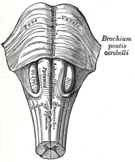
Babinski–Nageotte syndrome is an alternating brainstem syndrome. It occurs when there is damage to the dorsolateral or posterior lateral medulla oblongata, likely syphilitic in origin. Hence it is also called the alternating medulla oblongata syndrome.

Positron emission tomography–magnetic resonance imaging (PET–MRI) is a hybrid imaging technology that incorporates magnetic resonance imaging (MRI) soft tissue morphological imaging and positron emission tomography (PET) functional imaging.

Magnetic resonance imaging of the brain uses magnetic resonance imaging (MRI) to produce high quality two-dimensional or three-dimensional images of the brain and brainstem as well as the cerebellum without the use of ionizing radiation (X-rays) or radioactive tracers.

Jürgen Klaus Hennig is a German chemist and medical physicist. Internationally he is considered to be one of the pioneers of Magnetic Resonance Imaging for clinical diagnostics. He is the Scientific Director of the Department of Diagnostic Radiology and Chairman of the Magnetic Resonance Development and Application Center (MRDAC) at the University Medical Center Freiburg. In the year 2003 he was awarded the Max Planck Research Award in the category of Biosciences and Medicine.

Messoud Ashina is a Danish-Azerbaijani neurologist and neuroscientist. He is currently a Professor of Neurology at the University of Copenhagen and leads the Human Migraine Research Unit at the Danish Headache Center, Rigshospitalet. Ashina is also the Director of the Danish Knowledge Center on Headache Disorders and the past President of the International Headache Society. As of 2021, Ashina is ranked as the world's leading expert on headache disorders by Expertscape.

An MRI sequence in magnetic resonance imaging (MRI) is a particular setting of pulse sequences and pulsed field gradients, resulting in a particular image appearance.
Chandrasekharan Kesavadas is an Indian radiologist and a professor of radiology at the Sree Chitra Tirunal Institute for Medical Sciences and Technology. His research focus in the fields of magnetic resonance imaging, neuroradiology medical imaging informatics and brain Computer interface. He led a team of scientists who developed an imaging protocol for scanning intractable epilepsy, a computational software for detecting cortical dysplasia and novel neuroimaging systems for weighted imaging of brain cancer. His studies have been documented by way of a number of articles and ResearchGate, an online repository of scientific articles has listed 275 of them. The Department of Biotechnology of the Government of India awarded him the National Bioscience Award for Career Development, one of the highest Indian science awards, for his contributions to biosciences, in 2009. The award orations delivered by him include the 2010 edition of the M. N. Sen Oration of the Indian Council of Medical Research.
Denis Le Bihan is a medical doctor, physicist, member of the Institut de France, member of the French Academy of Technologies and director since 2007 of NeuroSpin, an institution of the Atomic Energy and Alternative Energy Commission (CEA) in Saclay, dedicated to the study of the brain by magnetic resonance imaging (MRI) with a very high magnetic field. Denis Le Bihan has received international recognition for his outstanding work, introducing new imaging methods, particularly for the study of the human brain, as evidenced by the many international awards he has received, such as the Gold Medal of the International Society of Magnetic Resonance in Medicine (2001), the coveted Lounsbery Prize, the Louis D. Prize from the Institut de France and the prestigious Honda Prize (2012). His work has focused on the introduction, development and application of highly innovative methods, notably diffusion MRI.
References
- ↑ Nielsen, Steffen Bang (3 June 2008). "MR-afdeling vil finde kræft med ny scanningsteknik" (in Danish). Archived from the original on 2 October 2011. Retrieved 20 November 2010.
- ↑ "Flere kan reddes efter blodprop" (in Danish). TV 2 Nyhederne. 12 September 2008. Retrieved 20 November 2010.
| General | |
|---|---|
| National libraries | |
| Scientific databases | |