Related Research Articles
Gray's Anatomy is a reference book of human anatomy written by Henry Gray, illustrated by Henry Vandyke Carter and first published in London in 1858. It has had multiple revised editions and the current edition, the 42nd, remains a standard reference, often considered "the doctors' bible".

The palate is the roof of the mouth in humans and other mammals. It separates the oral cavity from the nasal cavity. A similar structure is found in crocodilians, but in most other tetrapods, the oral and nasal cavities are not truly separated. The palate is divided into two parts, the anterior, bony hard palate and the posterior, fleshy soft palate.

A joint or articulation is the connection made between bones, ossicles, or other hard structures in the body which link an animal's skeletal system into a functional whole. They are constructed to allow for different degrees and types of movement. Some joints, such as the knee, elbow, and shoulder, are self-lubricating, almost frictionless, and are able to withstand compression and maintain heavy loads while still executing smooth and precise movements. Other joints such as sutures between the bones of the skull permit very little movement in order to protect the brain and the sense organs. The connection between a tooth and the jawbone is also called a joint, and is described as a fibrous joint known as a gomphosis. Joints are classified both structurally and functionally.

The inguinal canal is a passage in the anterior abdominal wall on each side of the body, which in males, convey the spermatic cords and in females, the round ligament of the uterus. The inguinal canals are larger and more prominent in males.
Gray's Anatomy is an 80-minute concert film directed by Steven Soderbergh in 1996 involving a dramatized monologue by actor/writer Spalding Gray. The title is taken from the classic human anatomy textbook Gray's Anatomy, originally written by Henry Gray in 1858. It was shot in ten days in late January 1996 during a break Soderbergh had from post-production on his previous film, Schizopolis.

Henry Gray was a British anatomist and surgeon most notable for publishing the book Gray's Anatomy. He was elected a Fellow of the Royal Society (FRS) at the age of 25.

In the human body, the lateral thoracic artery is a blood vessel that supplies oxygenated blood to approximately one-third of the lateral structures of the thorax and breast.

The external anal sphincter is an oval tube of skeletal muscle fibers. Distally, it is adherent to the skin surrounding the margin of the anus. It exhibits a resting state of tonical contraction and also contracts during the bulbospongiosus reflex.

The semilunar hiatus is a crescent-shaped/semicircular/ curved slit/groove upon the lateral wall of the nasal cavity at the middle nasal meatus just inferior to the ethmoidal bulla. It is the location of the openings for the frontal sinus, maxillary sinus, and anterior ethmoidal sinus. It is bounded inferiorly and anteriorly by the sharp concave margin of the uncinate process of the ethmoid bone, superiorly by the ethmoidal bulla, and posteriorly by the ethmoidal process of the inferior nasal concha. It leads into the ethmoidal infundibulum; it marks the medial limit of the ethmoidal infundibulum.
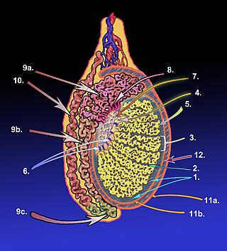
The tunica albuginea is a dense, blue-white layer of fibrous tissue surrounding the testis. It is the middle of three envelopes forming the capsule of the testis; it is deep to the visceral layer of tunica vaginalis, and superficial to the tunica vasculosa testis.

The sigmoid arteries are 2–5 branches of the inferior mesenteric artery that are distributed to the distal descending colon and the sigmoid colon.

The right colic artery is an artery of the abdomen, a branch of the superior mesenteric artery supplying the ascending colon. It divides into two terminal branches - an ascending branch and a descending branch - which form anastomoses with the middle colic artery, and ileocolic artery (respectively).

The posterior ethmoidal artery is an artery of the head which arises from the ophthalmic artery to supply the posterior ethmoidal air cells, and the meninges. It is smaller than the anterior ethmoidal artery.

General visceral efferent fibers (GVE), visceral efferents or autonomic efferents are the efferent nerve fibers of the autonomic nervous system that provide motor innervation to smooth muscle, cardiac muscle, and glands through postganglionic varicosities.

The sphenoidal spine is a downwardly directed process at the apex of the great wings of the sphenoid bone that serves as the origin of the sphenomandibular ligament.

The femoral ring is the opening at the proximal, abdominal end of the femoral canal, and represents the base of the conically-shaped femoral canal. The femoral ring is oval-shaped, with its long diameter being directed transversely and measuring about 1.25 cm. The opening of the femoral ring is filled in by extraperitoneal fat, forming the femoral septum.
The supraorbital artery is a branch of the ophthalmic artery. It passes anteriorly within the orbit to exit the orbit through the supraorbital foramen or notch alongside the supraorbital nerve, splitting into two terminal branches which go on to form anastomoses with arteries of the head.
The nerve to the stapedius is a branch of the facial nerve which innervates the stapedius muscle. It arises from the CN VII within the facial canal, opposite the pyramidal eminence. It passes through a small canal in this eminence to reach the stapedius muscle.

The aditus to mastoid antrum is a large, irregular opening upon the posterior wall of the tympanic cavity by which the mastoid antrum communicates with the epitympanic recess of the tympanic cavity. The walls of the antrum are lined by mucosa which is continuous with that lining the mastoid cells and tympanic cavity.

A nerve fascicle is a bundle of nerve fibers belonging to a nerve in the peripheral nervous system. A nerve fascicle is also called a fasciculus, as is a nerve tract in the central nervous system.
References
- ↑ "II. Osteology. 5d. The Interior of the Skull. Gray, Henry. 1918. Anatomy of the Human Body". Gray's Anatomy. Retrieved 2019-09-19.