
The central nervous system (CNS) is the part of the nervous system consisting primarily of the brain and spinal cord. The CNS is so named because it integrates the received information and coordinates and influences the activity of all parts of the bodies of bilaterally symmetric animals—i.e., all multicellular animals except sponges and radially symmetric animals such as jellyfish—and it contains the majority of the nervous system. The CNS also includes the retina and the optic nerve, as well as the olfactory nerves and olfactory epithelium as parts of the CNS, synapsing directly on brain tissue without intermediate ganglia. As such, the olfactory epithelium is the only central nervous tissue in direct contact with the environment, which opens up for therapeutic treatments. The CNS is contained within the dorsal body cavity, with the brain housed in the cranial cavity and the spinal cord in the spinal canal. In vertebrates, the brain is protected by the skull, while the spinal cord is protected by the vertebrae. The brain and spinal cord are both enclosed in the meninges. Within the CNS, the interneuronal space is filled with a large amount of supporting non-nervous cells called neuroglia or glia from the Greek for "glue".

The optic chiasm, or optic chiasma, is the part of the brain where the optic nerves cross. It is located at the bottom of the brain immediately inferior to the hypothalamus. The optic chiasm is found in all vertebrates, although in cyclostomes, it is located within the brain.
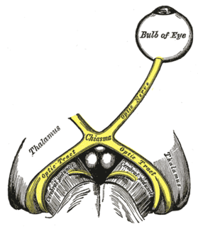
The optic nerve, also known as cranial nerve II, or simply as CN II, is a paired cranial nerve that transmits visual information from the retina to the brain. In humans, the optic nerve is derived from optic stalks during the seventh week of development and is composed of retinal ganglion cell axons and glial cells; it extends from the optic disc to the optic chiasma and continues as the optic tract to the lateral geniculate nucleus, pretectal nuclei, and superior colliculus.
Articles related to anatomy include:

The brainstem is the posterior part of the brain, continuous with the spinal cord. In the human brain the brainstem is composed of the midbrain, the pons, and the medulla oblongata. The midbrain is continuous with the thalamus of the diencephalon through the tentorial notch, and sometimes the diencephalon is included in the brainstem.
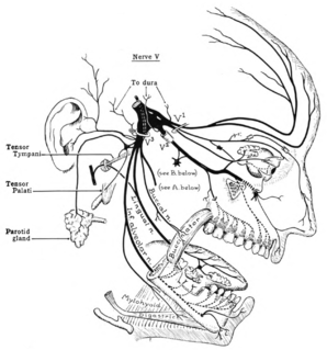
The trigeminal nerve (the fifth cranial nerve, or simply CN V) is a nerve responsible for sensation in the face and motor functions such as biting and chewing; it is the most complex of the cranial nerves. Its name ("trigeminal" = tri-, or three, and - geminus, or twin: thrice-twinned) derives from the fact that each of the two nerves (one on each side of the pons) has three major branches: the ophthalmic nerve (V1), the maxillary nerve (V2), and the mandibular nerve (V3). The ophthalmic and maxillary nerves are purely sensory, whereas the mandibular nerve supplies motor as well as sensory (or "cutaneous") functions. Adding to the complexity of this nerve is the fact that autonomic nerve fibers as well as special sensory fibers (taste) are contained within it.

The visual system comprises the sensory organ and the part of the central nervous system which gives organisms the ability to process visual detail as sight, as well as enabling the formation of several non-image photo response functions. It detects and interprets information from visible light to "build a representation" of the surrounding environment. The visual system carries out a number of complex tasks, including the reception of light and the formation of monocular representations, the neural mechanisms underlying stereo vision, the identification and categorization of visual objects, assessing distances to and between objects, motion perception, guiding body movements in relation to the objects seen, colour vision, and more. The psychological side of visual information procesing is known as visual perception, a lack of which is called blindness. Non-image forming visual functions, independent of visual perception, include the pupillary light reflex (PLR) and circadian photoentrainment.

The lateral geniculate nucleus is a relay center in the thalamus for the visual pathway. It receives a major sensory input from the retina. The LGN is the main central connection for the optic nerve to the occipital lobe, particularly the primary visual cortex. In humans, each LGN has six layers of neurons alternating with optic fibers.

The midbrain or mesencephalon is the forward-most portion of the brainstem and is associated with vision, hearing, motor control, sleep and wakefulness, arousal (alertness), and temperature regulation. The name comes from the Greek mesos, "middle", and enkephalos, "brain".

The internal capsule is a white matter structure situated in the inferomedial part of each cerebral hemisphere of the brain. It carries information past the basal ganglia, separating the caudate nucleus and the thalamus from the putamen and the globus pallidus. The internal capsule contains both ascending and descending axons, going to and coming from the cerebral cortex. It also separates the caudate nucleus and the putamen in the dorsal striatum, a brain region involved in motor and reward pathways.

The pupillary light reflex (PLR) or photopupillary reflex is a reflex that controls the diameter of the pupil, in response to the intensity (luminance) of light that falls on the retinal ganglion cells of the retina in the back of the eye, thereby assisting in adaptation of vision to various levels of lightness/darkness. A greater intensity of light causes the pupil to constrict, whereas a lower intensity of light causes the pupil to dilate. Thus, the pupillary light reflex regulates the intensity of light entering the eye. Light shone into one eye will cause both pupils to constrict.
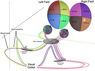
The optic radiation are axons from the neurons in the lateral geniculate nucleus to the primary visual cortex. The optic radiation receives blood through deep branches of the middle cerebral artery and posterior cerebral artery.
The optic tract is a part of the visual system in the brain. It is a continuation of the optic nerve that relays information from the optic chiasm to the ipsilateral lateral geniculate nucleus (LGN), pretectal nuclei, and superior colliculus.

The accommodation reflex is a reflex action of the eye, in response to focusing on a near object, then looking at a distant object, comprising coordinated changes in vergence, lens shape (accommodation) and pupil size. It is dependent on cranial nerve II, superior centers (interneuron) and cranial nerve III. The change in the shape of the lens is controlled by the ciliary muscles inside the eye. Changes in contraction of the ciliary muscles alter the focal distance of the eye, causing nearer or farther images to come into focus on the retina; this process is known as accommodation. The reflex, controlled by the parasympathetic nervous system, involves three responses: pupil constriction, lens accommodation, and convergence.

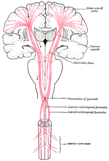
The corticobulbartract is a two-neuron white matter motor pathway connecting the motor cortex in the cerebral cortex to the medullary pyramids, which are part of the brainstem's medulla oblongata region, and are primarily involved in carrying the motor function of the non-oculomotor cranial nerves. The corticobulbar tract is one of the pyramidal tracts, the other being the corticospinal tract.
An upper motor neuron lesion occurs in the neural pathway above the anterior horn cell of the spinal cord or motor nuclei of the cranial nerves. Conversely, a lower motor neuron lesion affects nerve fibers traveling from the anterior horn of the spinal cord or the cranial motor nuclei to the relevant muscle(s).
The pretectal area, or pretectum, is a midbrain structure composed of seven nuclei and comprises part of the subcortical visual system. Through reciprocal bilateral projections from the retina, it is involved primarily in mediating behavioral responses to acute changes in ambient light such as the pupillary light reflex, the optokinetic reflex, and temporary changes to the circadian rhythm. In addition to the pretectum's role in the visual system, the anterior pretectal nucleus has been found to mediate somatosensory and nociceptive information.
The menace response is one of three forms of blink reflex. It is the reflex blinking that occurs in response to the rapid approach of an object. The reflex comprises blinking of the eyelids, in order to protect the eyes from potential damage, but may also include turning of the head, neck, or even the trunk away from the optical stimulus that triggers the reflex.

The visual pathway consists of structures that carry visual information from the retina to the brain. Lesions in that pathway cause a variety of visual field defects. In the visual system of human eye, the visual information processed by retinal photoreceptor cells travel in the following way:
Retina→Optic nerve→Optic chiasm →Optic tract→Lateral geniculate nucleus→Optic radiation→Primary and secondary visual cortices.