Assistive technology (AT) is a term for assistive, adaptive, and rehabilitative devices for people with disabilities and the elderly. Disabled people often have difficulty performing activities of daily living (ADLs) independently, or even with assistance. ADLs are self-care activities that include toileting, mobility (ambulation), eating, bathing, dressing, grooming, and personal device care. Assistive technology can ameliorate the effects of disabilities that limit the ability to perform ADLs. Assistive technology promotes greater independence by enabling people to perform tasks they were formerly unable to accomplish, or had great difficulty accomplishing, by providing enhancements to, or changing methods of interacting with, the technology needed to accomplish such tasks. For example, wheelchairs provide independent mobility for those who cannot walk, while assistive eating devices can enable people who cannot feed themselves to do so. Due to assistive technology, disabled people have an opportunity of a more positive and easygoing lifestyle, with an increase in "social participation," "security and control," and a greater chance to "reduce institutional costs without significantly increasing household expenses." In schools, assistive technology can be critical in allowing students with disabilities access the general education curriculum. Students who experience challenges writing or keyboarding, for example, can use voice recognition software instead. Assistive technologies assist people who are recovering from strokes and people who have abstained injuries that effect their daily tasks.

Photonics is a branch of optics that involves the application of generation, detection, and manipulation of light in form of photons through emission, transmission, modulation, signal processing, switching, amplification, and sensing. Photonics is closely related to quantum electronics, where quantum electronics deals with the theoretical part of it while photonics deal with its engineering applications. Though covering all light's technical applications over the whole spectrum, most photonic applications are in the range of visible and near-infrared light. The term photonics developed as an outgrowth of the first practical semiconductor light emitters invented in the early 1960s and optical fibers developed in the 1970s.

A smoke detector is a device that senses smoke, typically as an indicator of fire. Smoke detectors are usually housed in plastic enclosures, typically shaped like a disk about 150 millimetres (6 in) in diameter and 25 millimetres (1 in) thick, but shape and size vary. Smoke can be detected either optically (photoelectric) or by physical process (ionization). Detectors may use one or both sensing methods. Sensitive alarms can be used to detect and deter smoking in banned areas. Smoke detectors in large commercial and industrial buildings are usually connected to a central fire alarm system.

Electromyography (EMG) is a technique for evaluating and recording the electrical activity produced by skeletal muscles. EMG is performed using an instrument called an electromyograph to produce a record called an electromyogram. An electromyograph detects the electric potential generated by muscle cells when these cells are electrically or neurologically activated. The signals can be analyzed to detect abnormalities, activation level, or recruitment order, or to analyze the biomechanics of human or animal movement. Needle EMG is an electrodiagnostic medicine technique commonly used by neurologists. Surface EMG is a non-medical procedure used to assess muscle activation by several professionals, including physiotherapists, kinesiologists and biomedical engineers. In computer science, EMG is also used as middleware in gesture recognition towards allowing the input of physical action to a computer as a form of human-computer interaction.

Laser Doppler velocimetry, also known as laser Doppler anemometry, is the technique of using the Doppler shift in a laser beam to measure the velocity in transparent or semi-transparent fluid flows or the linear or vibratory motion of opaque, reflecting surfaces. The measurement with laser Doppler anemometry is absolute and linear with velocity and requires no pre-calibration.

A biosignal is any signal in living beings that can be continually measured and monitored. The term biosignal is often used to refer to bioelectrical signals, but it may refer to both electrical and non-electrical signals. The usual understanding is to refer only to time-varying signals, although spatial parameter variations are sometimes subsumed as well.
The mechanomyogram (MMG) is the mechanical signal observable from the surface of a muscle when the muscle is contracted. At the onset of muscle contraction, gross changes in the muscle shape cause a large peak in the MMG. Subsequent vibrations are due to oscillations of the muscle fibres at the resonance frequency of the muscle. The mechanomyogram is also known as the phonomyogram, acoustic myogram, sound myogram, vibromyogram or muscle sound.
Electro-optical sensors are electronic detectors that convert light, or a change in light, into an electronic signal. These sensors are able to detect electromagnetic radiation from the infrared up to the ultraviolet wavelengths. They are used in many industrial and consumer applications, for example:

Ultrasonic transducers and ultrasonic sensors are devices that generate or sense ultrasound energy. They can be divided into three broad categories: transmitters, receivers and transceivers. Transmitters convert electrical signals into ultrasound, receivers convert ultrasound into electrical signals, and transceivers can both transmit and receive ultrasound.
Targeted reinnervation enables amputees to control motorized prosthetic devices and to regain sensory feedback. The method was developed by Dr. Todd Kuiken at Northwestern University and Rehabilitation Institute of Chicago and Dr. Gregory Dumanian at Northwestern University Division of Plastic Surgery.

Tensiomyography(TMG) is a measuring method for detection of skeletal muscles' contractile properties. Tensiomyography assesses muscle mechanical response based on radial muscle belly displacement induced by a single electrical stimulus. It is performed using the TMG S2 system. A tensiomyography measurement instrument includes an electrical stimulator and data acquisition subunit (1), a digital sensor (2), a tripod with manipulating hand (3), and muscle electrodes (4) that work with an essential software interface installed on a PC.

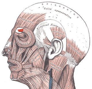
Facial electromyography (fEMG) refers to an electromyography (EMG) technique that measures muscle activity by detecting and amplifying the tiny electrical impulses that are generated by muscle fibers when they contract.

Magnetomyography (MMG) is a technique for mapping muscle activity by recording magnetic fields produced by electrical currents occurring naturally in the muscles, using arrays of SQUIDs. It has a better capability than electromyography for detecting slow or direct currents. The magnitude of the MMG signal is in the scale of pico (10−12) to femto (10−15) Tesla (T). Miniaturizing MMG offers a prospect to modernize the bulky SQUID to wearable miniaturized magnetic sensors.

A myograph is any device used to measure the force produced by a muscle when under contraction. Such a device is commonly used in myography, the study of the velocity and intensity of muscular contraction.
Clinical Electrophysiological Testing is based on techniques derived from electrophysiology used for the clinical diagnosis of patients. There are many processes that occur in the body which produce electrical signals that can be detected. Depending on the location and the source of these signals, distinct methods and techniques have been developed to properly target them.

Neuromechanics is an interdisciplinary field that combines biomechanics and neuroscience to understand how the nervous system interacts with the skeletal and muscular systems to enable animals to move. In a motor task, like reaching for an object, neural commands are sent to motor neurons to activate a set of muscles, called muscle synergies. Given which muscles are activated and how they are connected to the skeleton, there will be a corresponding and specific movement of the body. In addition to participating in reflexes, neuromechanical process may also be shaped through motor adaptation and learning.

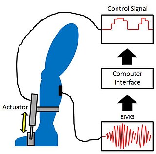
Proportional myoelectric control can be used to activate robotic lower limb exoskeletons. A proportional myoelectric control system utilizes a microcontroller or computer that inputs electromyography (EMG) signals from sensors on the leg muscle(s) and then activates the corresponding joint actuator(s) proportionally to the EMG signal.
Electrodiagnosis (EDX) is a method of medical diagnosis that obtains information about diseases by passively recording the electrical activity of body parts or by measuring their response to external electrical stimuli. The most widely used methods of recording spontaneous electrical activity are various forms of electrodiagnostic testing (electrography) such as electrocardiography (ECG), electroencephalography (EEG), and electromyography (EMG). Electrodiagnostic medicine is a medical subspecialty of neurology, clinical neurophysiology, cardiology, and physical medicine and rehabilitation. Electrodiagnostic physicians apply electrophysiologic techniques, including needle electromyography and nerve conduction studies to diagnose, evaluate, and treat people with impairments of the neurologic, neuromuscular, and/or muscular systems. The provision of a quality electrodiagnostic medical evaluation requires extensive scientific knowledge that includes anatomy and physiology of the peripheral nerves and muscles, the physics and biology of the electrical signals generated by muscle and nerve, the instrumentation used to process these signals, and techniques for clinical evaluation of diseases of the peripheral nerves and sensory pathways.
Robotic prosthesis control is a method for controlling a prosthesis in such a way that the controlled robotic prosthesis restores a biologically accurate gait to a person with a loss of limb. This is a special branch of control that has an emphasis on the interaction between humans and robotics.
An implantable myoelectric sensor (IMES) is a sensor implanted in or near a muscular region of the body in order to read the electric outputs of the muscles. This allows the device to measure the exact degree of activation of the muscle. This device is primarily used in disabled individuals as a detection module that feeds information regarding movement to externally powered and controlled prosthetics. The IMES was invented by Dr. Richard F. ff. Weir of the Rehabilitation Institute of Chicago and Dr. Philip Troyk of the Illinois Institute of Technology, with the first experimental studies performed by Jack F. Schorsch of the Rehabilitation Institute of Chicago.


