Orientation column

Last updated

Orientation columns are organized regions of neurons that are excited by visual line stimuli of varying angles. These columns are located in the primary visual cortex (V1) and span multiple cortical layers. The geometry of the orientation columns are arranged in slabs that are perpendicular to the surface of the primary visual cortex. [1] [2]

Contents

The primary visual cortex (V1) is located in the occipital lobe. This is the region where orientation columns are found. Visualcortex.gif
The primary visual cortex (V1) is located in the occipital lobe. This is the region where orientation columns are found.

History

In 1958, David Hubel and Torsten Wiesel discovered cells in the visual cortex that had orientation selectivity. This was found through an experiment by giving a cat specific visual stimuli and measuring the corresponding excitation of the neurons in striate cortex (V1). The experimental set up was of a slide projector, a cat, electrodes, and an electrode monitor. They discovered this orientation selectivity when changing slides on the projector. The act of changing the slides produced a faint shadow line across the projector, and excited the neuron they were measuring. At the time of this experiment it was not conclusive that these orientation selective cells were in a "columnar" structure but the possibility of this structure was considered by research conducted by Vernon Mountcastle in 1956 about the topographic properties of the somatosensory system. [3] [4] [5]

In 1974 Hubel and Wiesel wrote a paper about the geometry of orientation columns. They recorded 1410 cells in 45 penetrations into the striate cortex. Through this 1-dimensional technique they conceptualized that the orientation columns are not columns but slabs. [1] In 1985, Gary Blasdel discovered a technique to visualize these orientation columns in 2D. His technique used photodiodes to detect optical changes in the visual cortex with the metabolic marker, 2-deoxyglucose, which labels active neurons. This confirmed Hubel and Wiesel's studies and also brought to light the swirls and pinwheel formations in the striate cortex. [4] [6]

Hubel and Wiesel received the Nobel Prize in Physiology and Medicine in 1981 for their contributions to our knowledge of the development of the visual system. [4]

Physiology

Orientation columns are located in the primary visual cortex also known as the striate cortex. These orientation columns are not cylindrical in shape as the word column implies but are flat slabs that are parallel to each other. The slabs are perpendicular to the surface of the visual cortex and are lined up similar to slices of bread. These neurons are highly discriminatory for visual orientations and their motion. [1] [7]

Most of the cells in orientation columns are complex cells. Complex cells will respond to a properly orientated line in any location of the receptive field, whereas simple cells have a narrower receptive field where a properly oriented line will excite it. Simple cells have distinct subdivisions of excitatory and inhibitory regions. It is proposed that complex cells receive input from many simple cells, which explains why the complex cells have a slightly wider receptive field. [8] [9]

There are possible biological advantages to the highly ordered structures of orientation columns. The first possible advantage is that orientation selectivity may be intensified with lateral inhibition from neighboring cells of a slightly different preferred orientation. This would provide an efficient system for wiring between the striate cortex and the lateral geniculate nucleus (LGN). [10] The second possible advantage is the ordered structure aids in development, by guaranteeing all orientations are represented throughout the visual field with minimal redundancy and no deficiencies. The third possible advantage is that if columns with similar orientation selectivity are close together, fewer afferents from the LGN are needed. This allows for efficient wiring. So by removing a few LGN inputs and adding a few, the orientation selectivity can be changed marginally. [1]

Ocular dominance columns are also found in the striate cortex. These columns were found to prefer crossing iso-orientation lines perpendicularly. During microelectrode experiments, it is normal to see penetrations where eye dominance changes between the contralateral eye and ipsilateral eye but this does not interrupt the orientation sequence. [7]

Preferred orientation

Recently, studies involving human models were conducted with high-field fMRI. These studies demonstrated the existence of orientation preference in humans and showed similarities to the studies conducted with monkey models. It was found in these models that there was an over-representation of the 90° orientation preference. This corresponds to a bias towards processing vertical visual stimuli with horizontal movement. This bears resemblance to the oblique effect where there is a deficiency in perception to oblique contours (45° and 90°). [11]

Pinwheels

Pinwheel formations in the primary visual cortex with singularities in the center. Each color represents an orientation column of a specific line phase. Adapted image from fMRI studies. VisualSingularity2.jpg
Pinwheel formations in the primary visual cortex with singularities in the center. Each color represents an orientation column of a specific line phase. Adapted image from fMRI studies.

Using 2D optical techniques, pinwheel formations (also known as whorls) of orientation columns were discovered. Pinwheels are the location where multiple orientation columns converge. Orientation columns are organized radially around a point known as a singularity. The arrangement, around the singularity, can be observed to be in both a counter-clockwise or clockwise fashion. [11] It is suggested that an artifact of the optical recordings may cause these singularities. Limitations in the resolution of the optical technique may be to explain for these singularities. [4]

Fractures

Fractures are breaks in the sequence of orientation selectivity from microelectrode studies. In these studies the fractures occur randomly during trials. [1] Optical methods in trying to explain why these fractures occur, have had limited success. [4]

Development

Orientation maps in monkeys are innately determined at birth. Like other parts of the brain, the visual cortex goes through a critical period where the visual environment can change the orientation maps due to its plastic nature during this period. Visual deprivation during this period will cause a deterioration of these innate connections. [12] Also if the visual environment is confined to only vertical or horizontal lines during this critical period the distribution of the preferred orientation of cells in the striate cortex become abnormal. This is probably due to cells maturing their preferred orientation to that of the most common type of visual stimulus. [13]

Modeling

Hebbian development of a multilayer neural network

A multilayer neural network model by Linsker, having local connections from each cell layer to the next, whose connection strengths develop according to a Hebbian rule, generates orientation-selective cells and orientation columns. [14] The resulting columnar arrangement contains fractures and "pinwheel" singularities of the same types as those found experimentally.

Moire interference

An example of a Moire interference pattern. The offset of the two lattices creates a dipole of retinal ganglion cells. This dipole is orientated in various directions that correspond to a specific orientation. Moire-(quadrat)-1.png
An example of a Moiré interference pattern. The offset of the two lattices creates a dipole of retinal ganglion cells. This dipole is orientated in various directions that correspond to a specific orientation.

A highly debated [15] [16] model for the origin of orientation maps is Moiré interference from retinal ganglion cells (RGCs). [17] The ideal case takes two layers of perfect hexagonal lattices of the on-center and off-center receptive fields of the RGCs. These two layers are superimposed on each other with an angled offset that produces a periodic interference pattern. This pattern produces dipoles of these RGCs that have a preferred orientation scattered throughout the visual field. This mosaic produces periodic map of preferred orientation that fulfills all orientations with regularity. Cortical inputs from this mosaic of RGCs through the LGN can explain the origin of the orientation maps in the visual cortex.

Further research

Orientation scotomas

The theory of Moire interference patterns governing the orientation map predicts the existence of orientation scotomas. This is because the lattice of the RGCs are not perfectly hexagonal and therefore, at some locations, representation of specific orientations will be missing. Currently there is research that is testing this hypothesis by "mapping human orientation discrimination thresholds of very small stimuli in the far periphery." [17]

Further reading

See also

Related Research Articles

Visual cortex Region of the brain that processes visual information

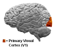
The visual cortex of the brain is the area of the cerebral cortex that processes visual information. It is located in the occipital lobe. Sensory input originating from the eyes travels through the lateral geniculate nucleus in the thalamus and then reaches the visual cortex. The area of the visual cortex that receives the sensory input from the lateral geniculate nucleus is the primary visual cortex, also known as visual area 1 (V1), Brodmann area 17, or the striate cortex. The extrastriate areas consist of visual areas 2, 3, 4, and 5.

Visual system Body parts responsible for sight

The visual system comprises the sensory organ and parts of the central nervous system which gives organisms the sense of sight as well as enabling the formation of several non-image photo response functions. It detects and interprets information from the optical spectrum perceptible to that species to "build a representation" of the surrounding environment. The visual system carries out a number of complex tasks, including the reception of light and the formation of monocular neural representations, colour vision, the neural mechanisms underlying stereopsis and assessment of distances to and between objects, the identification of particular object of interest, motion perception, the analysis and integration of visual information, pattern recognition, accurate motor coordination under visual guidance, and more. The neuropsychological side of visual information processing is known as visual perception, an abnormality of which is called visual impairment, and a complete absence of which is called blindness. Non-image forming visual functions, independent of visual perception, include the pupillary light reflex (PLR) and circadian photoentrainment.

Torsten Wiesel

Torsten Nils Wiesel is a Swedish neurophysiologist. Together with David H. Hubel, he received the 1981 Nobel Prize in Physiology or Medicine, for their discoveries concerning information processing in the visual system; the prize was shared with Roger W. Sperry for his independent research on the cerebral hemispheres.

David H. Hubel

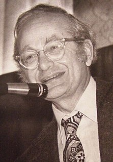
David Hunter Hubel was a Canadian American neurophysiologist noted for his studies of the structure and function of the visual cortex. He was co-recipient with Torsten Wiesel of the 1981 Nobel Prize in Physiology or Medicine, for their discoveries concerning information processing in the visual system. For much of his career, Hubel was the John Franklin Enders University Professor of Neurobiology at Harvard Medical School. In 1978, Hubel and Wiesel were awarded the Louisa Gross Horwitz Prize from Columbia University.In 1983, Hubel received the Golden Plate Award of the American Academy of Achievement.

A cortical column, also called hypercolumn, macrocolumn, functional column or sometimes cortical module, is a group of neurons in the cortex of the brain that can be successively penetrated by a probe inserted perpendicularly to the cortical surface, and which have nearly identical receptive fields. Neurons within a minicolumn (microcolumn) encode similar features, whereas a hypercolumn "denotes a unit containing a full set of values for any given set of receptive field parameters". A cortical module is defined as either synonymous with a hypercolumn (Mountcastle) or as a tissue block of multiple overlapping hypercolumns.

According to Alonso and Chen (2008),

The receptive field is a portion of sensory space that can elicit neuronal responses when stimulated. The sensory space can be defined in a single dimension, two dimensions or multiple dimensions. The neuronal response can be defined as firing rate or include also subthreshold activity.

Neuronal tuning refers to the hypothesized property of brain cells by which they selectively represent a particular type of sensory, association, motor, or cognitive information. Some neuronal responses have been hypothesized to be optimally tuned to specific patterns through experience. Neuronal tuning can be strong and sharp, as observed in primary visual cortex, or weak and broad, as observed in neural ensembles. Single neurons are hypothesized to be simultaneously tuned to several modalities, such as visual, auditory, and olfactory. Neurons hypothesized to be tuned to different signals are often hypothesized to integrate information from the different sources. In computational models called neural networks, such integration is the major principle of operation. The best examples of neuronal tuning can be seen in the visual, auditory, olfactory, somatosensory, and memory systems, although due to the small number of stimuli tested the generality of neuronal tuning claims is still an open question.

Ocular dominance columns are stripes of neurons in the visual cortex of certain mammals that respond preferentially to input from one eye or the other. The columns span multiple cortical layers, and are laid out in a striped pattern across the surface of the striate cortex (V1). The stripes lie perpendicular to the orientation columns.

The neocognitron is a hierarchical, multilayered artificial neural network proposed by Kunihiko Fukushima in 1979. It has been used for handwritten character recognition and other pattern recognition tasks, and served as the inspiration for convolutional neural networks.

Simple cell

A simple cell in the primary visual cortex is a cell that responds primarily to oriented edges and gratings. These cells were discovered by Torsten Wiesel and David Hubel in the late 1950s.

Complex cells can be found in the primary visual cortex (V1), the secondary visual cortex (V2), and Brodmann area 19 (V3).

Colour centre

The colour centre is a region in the brain primarily responsible for visual perception and cortical processing of colour signals received by the eye, which ultimately results in colour vision. The colour centre in humans is thought to be located in the ventral occipital lobe as part of the visual system, in addition to other areas responsible for recognizing and processing specific visual stimuli, such as faces, words, and objects. Many functional magnetic resonance imaging (fMRI) studies in both humans and macaque monkeys have shown colour stimuli to activate multiple areas in the brain, including the fusiform gyrus and the lingual gyrus. These areas, as well as others identified as having a role in colour vision processing, are collectively labelled visual area 4 (V4). The exact mechanisms, location, and function of V4 are still being investigated.

Hypercomplex cell

A hypercomplex cell is a type of visual processing neuron in the mammalian cerebral cortex. Initially discovered by David Hubel and Torsten Wiesel in 1965, hypercomplex cells are defined by the property of end-stopping, which is a decrease in firing strength with increasingly larger stimuli. The sensitivity to stimulus length is accompanied by selectivity for the specific orientation, motion, and direction of stimuli. For example, a hypercomplex cell may only respond to a line at 45˚ that travels upward. Elongating the line would result in a proportionately weaker response. Ultimately, hypercomplex cells can provide a means for the brain to visually perceive corners and curves in the environment by identifying the ends of a given stimulus.

Feature detection is a process by which the nervous system sorts or filters complex natural stimuli in order to extract behaviorally relevant cues that have a high probability of being associated with important objects or organisms in their environment, as opposed to irrelevant background or noise.

Monocular deprivation is an experimental technique used by neuroscientists to study central nervous system plasticity. Generally, one of an animal's eyes is sutured shut during a period of high cortical plasticity. This manipulation serves as an animal model for amblyopia, a permanent deficit in visual sensation not due to abnormalities in the eye.

Oblique effect is the name given to the relative deficiency in perceptual performance for oblique contours as compared to the performance for horizontal or vertical contours.

Visual tilt effects

Due to the effect of a spatial context or temporal context, the perceived orientation of a test line or grating pattern can appear tilted away from its physical orientation. The tilt illusion (TI) is the phenomenon that the perceived orientation of a test line or grating is altered by the presence of surrounding lines or grating with a different orientation. And the tilt aftereffect (TAE) is the phenomenon that the perceived orientation is changed after prolonged inspection of another oriented line or grating.

Binocular neurons are neurons in the visual system that assist in the creation of stereopsis from binocular disparity. They have been found in the primary visual cortex where the initial stage of binocular convergence begins. Binocular neurons receive inputs from both the right and left eyes and integrate the signals together to create a perception of depth.

Russell L. De Valois

Russell L. De Valois was an American scientist recognized for his pioneering research on spatial and color vision.

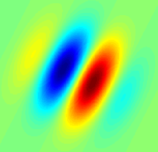
Orientation selectivity is expressed by cells within the visual cortex, when such cells increase impulse or signal activity for specific oriented degree of shape presented within the visual field. Orientation selectivity can also be expressed by simple cells if the orientation of a stimulus is orthogonal to the preferred degree of orientation, which results in the inhibition of impulse activity.

References

  1. 1 2 3 4 5 Hubel, D. H., & Wiesel, T. N. (1974). SEQUENCE REGULARITY AND GEOMETRY OF ORIENTATION COLUMNS IN MONKEY STRIATE CORTEX. [Article]. Journal of Comparative Neurology, 158(3), 267-294.
  2. Hubel, D. H., & Wiesel, T. N. (1968). RECEPTIVE FIELDS AND FUNCTIONAL ARCHITECTURE OF MONKEY STRIATE CORTEX. Journal of Physiology-London, 195(1), 215-&.
  3. Hubel, D. H., & Wiesel, T. N. (1959). Receptive Fields of Single Neurones in the Cat's Striate Cortex. [Article]. Journal of Physiology, 148, 574-591.
  4. 1 2 3 4 5 Hubel, D. H., & Wiesel, T. N. (2005). Brain and Visual Perception. New York: Oxford Press.
  5. Mountcastle, V. B. (1956). Modality and Topographic Properties of Single Neurons of Cat's Somatic Sensory Cortex. [Article]. Journal of Neurophysiology, 20(4), 408-435.
  6. Blasdel G. G., & Salama G. (1986). Voltage-sensitive dyes reveal a modular organization in monkey striate cortex. [Article]. Nature, 321, 579-585.
  7. 1 2 Hubel, D. H., & Wiesel, T. N. (1977). Ferrier Lecture: Function architecture of macaque monkey visual cortex. [Typescript]. Proc. R. Soc. Lond., 198, 1-59.
  8. Hubel, D. H., & Wiesel, T. N. (1962). Receptive fields, binocular interaction and functional architecture in the cat's visual cortex. [Article]. Journal of Physiology, 160, 106-154.
  9. Hubel, D. H. (1995). Eye, Brain and Vision. Scientific American Library.
  10. Blakemore, C., & Tobin, E. A. (1972). Lateral inhibition between orientation detectors in the cat's visual cortex. [Article]. Exp. Brain Res., 15, 439-440.
  11. 1 2 3 Yacoub, E., & Harel, N., & Ugurbil, K. (2008). High-Field fMRI unveils orientation columns in humans. [Article]. Proc Natl Acad Sci, 105, 10607-10612.
  12. Hubel, D. H., & Wiesel, T. N. (1974). Ordered Arrangement of Orientation Columns in Monkeys Lacking Visual Experience. [Article]. Journal of Comparative Neurology, 158, 307-318.
  13. Blakemore, C., Cooper, G. F. (1970). Development of the brain depends on the visual environment. Nature, 228, 477-478.
  14. Linsker R. (1986). From basic network principles to neural architecture (series of three papers). PNAS 83, 7508-7512, 8390-8394, 8779-8783. doi:10.1073/pnas.83.19.7508; doi:10.1073/pnas.83.21.8390; doi:10.1073/pnas.83.22.8779 .
  15. Schottdorf M., Eglen S. J., Wolf F. & Keil W. (2014). Can Retinal Ganglion Cell Dipoles Seed Iso-Orientation Domains in the Visual Cortex? PLoS ONE 9(1), e86139. doi:10.1371/journal.pone.0086139.
  16. Hore, V. R. A., Troy, J. B., & Eglen, S. J. (2012). Parasol cell mosaics are unlikely to drive the formation of structured orientation maps in primary visual cortex. Visual Neuroscience, 29(6), 283–299. doi:10.1017/S0952523812000338.
  17. 1 2 Paik, S., & Ringach, D. L. (2011). Retinal origin of orientation maps in the visual cortex. Nature Neuroscience, 14(7), 919-925.