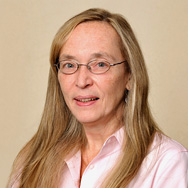
Pamela Robey

Last updated
Pamela Robey
Pamela Robey.jpg
Alma mater Susquehanna University (B.A.)
Catholic University of America (M.S., Ph.D.)
Scientific career
FieldsSkeletal cell biology
Institutions National Institute of Dental and Craniofacial Research
Thesis Studies on the Collagenous Component of a Tumor Basement Membrane  (1979)

Pamela Gehron Robey (born 1952) is an American cell biologist. She is a senior investigator in the skeletal biology section at the National Institute of Dental and Craniofacial Research.

Contents

Education

Robey received her B.A. in biology from Susquehanna University in 1974. She completed a M.S. in biochemistry in 1977 and a Ph.D. in cell biology in 1979 from the Catholic University of America. [1] Her Master's graduate thesis was titled The isolation, purification and characterization of A/J skin collagen and the effects of dexamethasone on skin collegen metabolism. [2] Her 1979 dissertation was titled Studies on the Collagenous Component of a Tumor Basement Membrane. [3] She did her post-doctoral work at the National Institute of Arthritis, Metabolism and Digestive Diseases on the role of defective phosphorylation of enzymes leading to lysosomal storage disease, and a staff fellowship in the National Eye Institute, where she studied retinal and ocular connective tissue diseases. [4]

Career

Robey joined NIDCR in 1983 and established reproducible methods for culturing human bone-forming cells, in order to study the development of mineralized matrix formation. In 1992, Robey was appointed chief of Skeletal Biology Section. Robey has served as a co-coordinator of the NIH Bone Marrow Stromal Cell Transplantation Center (2008-2013), and is currently the acting Scientific Director of the NIH Stem Cell Characterization Facility. She is a senior investigator in the skeletal biology section at NIDCR. [4] In early 2000s, she discovered dental stem cells. [5]

Research interests

Robey focuses on four main areas in skeletal cell biology:  1) determination of the characteristics and the biological properties of post-natal bone marrow stromal cells (BMSCs), a subset of which are multipotent skeletal stem cells (SSCs), able to recreate cartilage, bone, cells that support blood formation and fat cells in the marrow; 2) elucidation of the role of enzymatic matrix remodeling in the maintenance of SSC function; 3) characterization of the role that BMSCs/SSCs play in skeletal diseases; and, 4) development of techniques for cartilage and bone regeneration in human patients with skeletal defects.  In addition to using BMSCs for tissue engineering, Dr. Robey and her group also explore the potential of pluripotent stem cells to differentiate into cartilage and bone as another source of cells for skeletal regeneration. [4]

Related Research Articles

<span class="mw-page-title-main">Bone</span> Rigid organs that constitute part of the endoskeleton of vertebrates

A bone is a rigid organ that constitutes part of the skeleton in most vertebrate animals. Bones protect the various other organs of the body, produce red and white blood cells, store minerals, provide structure and support for the body, and enable mobility. Bones come in a variety of shapes and sizes and have complex internal and external structures. They are lightweight yet strong and hard and serve multiple functions.

<span class="mw-page-title-main">Osteoblast</span> Cells secreting extracellular matrix

Osteoblasts are cells with a single nucleus that synthesize bone. However, in the process of bone formation, osteoblasts function in groups of connected cells. Individual cells cannot make bone. A group of organized osteoblasts together with the bone made by a unit of cells is usually called the osteon.

The National Institute of Arthritis and Musculoskeletal and Skin Diseases (NIAMS) is one of the institutes and centers that make up the National Institutes of Health, an agency of the United States Department of Health and Human Services (HHS).

<span class="mw-page-title-main">Hematopoietic stem cell</span> Stem cells that give rise to other blood cells

Hematopoietic stem cells (HSCs) are the stem cells that give rise to other blood cells. This process is called haematopoiesis. In vertebrates, the first definitive HSCs arise from the ventral endothelial wall of the embryonic aorta within the (midgestational) aorta-gonad-mesonephros region, through a process known as endothelial-to-hematopoietic transition. In adults, haematopoiesis occurs in the red bone marrow, in the core of most bones. The red bone marrow is derived from the layer of the embryo called the mesoderm.

<span class="mw-page-title-main">Chondrocyte</span> Cell that makes up cartilage

Chondrocytes are the only cells found in healthy cartilage. They produce and maintain the cartilaginous matrix, which consists mainly of collagen and proteoglycans. Although the word chondroblast is commonly used to describe an immature chondrocyte, the term is imprecise, since the progenitor of chondrocytes can differentiate into various cell types, including osteoblasts.

<span class="mw-page-title-main">Fibrous dysplasia of bone</span> Medical condition

Fibrous dysplasia is a very rare nonhereditary genetic disorder where normal bone and marrow is replaced with fibrous tissue, resulting in formation of bone that is weak and prone to expansion. As a result, most complications result from fracture, deformity, functional impairment, pain, and the impingement of nerves. Disease occurs along a broad clinical spectrum ranging from mostly asymptomatic incidental lesions, to severe disabling disease. Disease can affect one bone (monostotic), multiple (polyostotic), or all bones (panostotic) and may occur in isolation or in combination with café au lait skin macules and hyperfunctioning endocrinopathies, termed McCune–Albright syndrome. More rarely, fibrous dysplasia may be associated with intramuscular myxomas, termed Mazabraud's syndrome. Fibrous dysplasia is very rare, and there is no known cure. While fibrous dysplasia is not itself a form of cancer, in severe cases it may undergo a malignant transformation into cancers such as osteosarcoma or chondrosarcoma, so some clinicians may regard it as precancerous rather than benign.

Stromal cells, or mesenchymal stromal cells, are differentiating cells found in abundance within bone marrow but can also be seen all around the body. Stromal cells can become connective tissue cells of any organ, for example in the uterine mucosa (endometrium), prostate, bone marrow, lymph node and the ovary. They are cells that support the function of the parenchymal cells of that organ. The most common stromal cells include fibroblasts and pericytes. The term stromal comes from Latin stromat-, "bed covering", and Ancient Greek στρῶμα, strôma, "bed".

<span class="mw-page-title-main">Sp7 transcription factor</span> Protein-coding gene in the species Homo sapiens

Transcription factor Sp7, also called osterix (Osx), is a protein that in humans is encoded by the SP7 gene. It is a member of the Sp family of zinc-finger transcription factors It is highly conserved among bone-forming vertebrate species It plays a major role, along with Runx2 and Dlx5 in driving the differentiation of mesenchymal precursor cells into osteoblasts and eventually osteocytes. Sp7 also plays a regulatory role by inhibiting chondrocyte differentiation maintaining the balance between differentiation of mesenchymal precursor cells into ossified bone or cartilage. Mutations of this gene have been associated with multiple dysfunctional bone phenotypes in vertebrates. During development, a mouse embryo model with Sp7 expression knocked out had no formation of bone tissue. Through the use of GWAS studies, the Sp7 locus in humans has been strongly associated with bone mass density. In addition there is significant genetic evidence for its role in diseases such as Osteogenesis imperfecta (OI).

Mesenchymal stem cells (MSCs) are multipotent cells found in multiple human adult tissues, including bone marrow, synovial tissues, and adipose tissues. Since they are derived from the mesoderm, they have been shown to differentiate into bone, cartilage, muscle, and adipose tissue. MSCs from embryonic sources have shown promise scientifically while creating significant controversy. As a result, many researchers have focused on adult stem cells, or stem cells isolated from adult humans that can be transplanted into damaged tissue.

Dental pulp stem cells (DPSCs) are stem cells present in the dental pulp, which is the soft living tissue within teeth. DPSCs can be collected from dental pulp by means of a non-invasive practice. It can be performed with an adult after simple extraction or to the young after surgical extraction of wisdom teeth. They are pluripotent, as they can form embryoid body-like structures (EBs) in vitro and teratoma-like structures that contained tissues derived from all three embryonic germ layers when injected in nude mice. DPSCs can differentiate in vitro into tissues that have similar characteristics to mesoderm, endoderm and ectoderm layers. They can differentiate into many cell types, such as odontoblasts, neural progenitors, osteoblasts, chondrocytes, and adipocytes. DPSCs were found to be able to differentiate into adipocytes and neural-like cells. DPSC differentiation into osteogenic lines is enhanced in 3D condition and hypoxia. These cells can be obtained from postnatal teeth, wisdom teeth, and deciduous teeth, providing researchers with a non-invasive method of extracting stem cells. The different cell populations, however, differ in certain aspects of their growth rate in culture, marker gene expression and cell differentiation, although the extent to which these differences can be attributed to tissue of origin, function or culture conditions remains unclear. As a result, DPSCs have been thought of as an extremely promising source of cells used in endogenous tissue engineering.

<span class="mw-page-title-main">Mesenchymal stem cell</span> Multipotent, non-hematopoietic adult stem cells present in multiple tissues

Mesenchymal stem cells (MSCs) also known as mesenchymal stromal cells or medicinal signaling cells, are multipotent stromal cells that can differentiate into a variety of cell types, including osteoblasts, chondrocytes, myocytes and adipocytes.

<span class="mw-page-title-main">A. Hari Reddi</span> Professor

A. Hari Reddi is a Distinguished Professor and holder of the Lawrence J. Ellison Endowed Chair in Musculoskeletal Molecular Biology at the University of California, Davis. He was previously the Virginia M. and William A. Percy Chair and Professor in Orthopaedic Surgery, Professor of Biological Chemistry, and Professor of Oncology at the Johns Hopkins University School of Medicine. Professor Reddi's research played an indispensable role in the identification, isolation and purification of bone morphogenetic proteins (BMPs) that are involved in bone formation and repair. The molecular mechanism of bone induction studied by Professor Reddi led to the conceptual advance in tissue engineering that morphogens in the form of metabologens bound to an insoluble extracellular matrix scaffolding act in collaboration to stimulate stem cells to form cartilage and bone. The Reddi laboratory has also made important discoveries unraveling the role of the extracellular matrix in bone and cartilage tissue regeneration and repair.

<span class="mw-page-title-main">Martha Somerman</span> American scientist

Martha J. Somerman is an internationally known researcher and educator in medicine, focusing on defining the key regulators controlling development, maintenance, and regeneration of dental, oral, and craniofacial tissues. She was 'Chief Lab of Laboratory of Oral Connective Tissue Biology (LOCTB) at the National Institutes of Health's National Institute of Arthritis and Musculoskeletal and Skin Diseases (NIAMS) and Director of the National Institute of Dental and Craniofacial Research (NIDCR), a part of the National Institutes of Health (NIH) located in Bethesda, Maryland.

Craniofacial regeneration refers to the biological process by which the skull and face regrow to heal an injury. This page covers birth defects and injuries related to the craniofacial region, the mechanisms behind the regeneration, the medical application of these processes, and the scientific research conducted on this specific regeneration. This regeneration is not to be confused with tooth regeneration. Craniofacial regrowth is broadly related to the mechanisms of general bone healing.

<span class="mw-page-title-main">Marian Young</span> American developmental biologist

Marian Frances Young is an American developmental biologist researching the function of extracellular matrix proteins in skeletal tissues. She is the deputy scientific director of the division of intramural research at the National Institute of Dental and Craniofacial Research.

Ophir David Klein is an American developmental biologist who specializes in pediatric medical genetics. Klein is Executive Director of Cedars-Sinai Guerin Children’s, Vice Dean for Children’s Services, Professor of Pediatrics, and the David and Meredith Kaplan Distinguished Chair in Children’s Health. He is also a professor of Orofacial Sciences and Pediatrics at UCSF.

Lynda Bonewald is a professor of anatomy, cell biology, physiology, and orthopaedic surgery and the founding director of the Indiana Center for Musculoskeletal Health (ICMH) at the Indiana University School of Medicine. She studies bone and the musculoskeletal system. She has served as president of the American Society for Bone and Mineral Research and the Association of Biomolecular Resource Facilities (1999-2000).

<span class="mw-page-title-main">Kelly Ten Hagen</span> American glycobiologist

Kelly Greig Ten Hagen is an American glycobiologist and head of the developmental glycobiology section at the National Institute of Dental and Craniofacial Research. She studies O-glycosylation regulation and its relationship to human disease.

<span class="mw-page-title-main">Tracey Rouault</span> American rheumatologist and physician-scientist

Tracey Ann Rouault is an American rheumatologist and physician-scientist who researches mammalian iron-sulfur proteins. Rouault is a senior investigator at the Eunice Kennedy Shriver National Institute of Child Health and Human Development and she heads the section on human iron metabolism.

<span class="mw-page-title-main">Hynda Kleinman</span> American cell biologist

Hynda K. Kleinman is an American cell biologist who was the chief of the cell biology section at the National Institute of Dental and Craniofacial Research from 1985 to 2006. She co-invented Matrigel.

References

  1. "Principal Investigators". NIH Intramural Research Program. Retrieved 2019-09-19.
  2. Robey, Pamela Gehron (1977). The isolation, purification and characterization of A/J skin collagen and the effects of dexamethasone on skin collegen metabolism. OCLC   4822988.
  3. Robey, Pamela Gehron (1980). Studies on the collagenous component of a tumor basement membrane. OCLC   10944705.
  4. 1 2 3 "Pamela Robey, Ph.D." National Institute of Dental and Craniofacial Research. Retrieved 2019-09-19.
  5. "The Quest to Understand Dental Stem Cells | National Institute of Dental and Craniofacial Research". 19 November 2018.
PD-icon.svg This article incorporates public domain material from websites or documents of the National Institutes of Health.