Related Research Articles
Hemicorporectomy is a radical surgery in which the body below the waist is amputated, transecting the lumbar spine. This removes the legs, the genitalia, urinary system, pelvic bones, anus, and rectum. It is a major procedure recommended only as a last resort for people with severe and potentially fatal illnesses such as osteomyelitis, tumors, severe traumas and intractable decubiti in, or around, the pelvis. By 2009, 66 cases had been reported in medical literature.

Internal bleeding is a loss of blood from a blood vessel that collects inside the body, and is not usually visible from the outside. It can be a serious medical emergency but the extent of severity depends on bleeding rate and location of the bleeding. Severe internal bleeding into the chest, abdomen, pelvis, or thighs can cause hemorrhagic shock or death if proper medical treatment is not received quickly. Internal bleeding is a medical emergency and should be treated immediately by medical professionals.

Airway management includes a set of maneuvers and medical procedures performed to prevent and relieve airway obstruction. This ensures an open pathway for gas exchange between a patient's lungs and the atmosphere. This is accomplished by either clearing a previously obstructed airway; or by preventing airway obstruction in cases such as anaphylaxis, the obtunded patient, or medical sedation. Airway obstruction can be caused by the tongue, foreign objects, the tissues of the airway itself, and bodily fluids such as blood and gastric contents (aspiration).

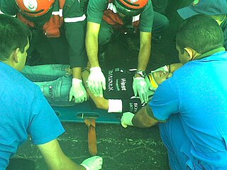
Major trauma is any injury that has the potential to cause prolonged disability or death. There are many causes of major trauma, blunt and penetrating, including falls, motor vehicle collisions, stabbing wounds, and gunshot wounds. Depending on the severity of injury, quickness of management, and transportation to an appropriate medical facility may be necessary to prevent loss of life or limb. The initial assessment is critical, and involves a physical evaluation and also may include the use of imaging tools to determine the types of injuries accurately and to formulate a course of treatment.

A bone fracture is a medical condition in which there is a partial or complete break in the continuity of any bone in the body. In more severe cases, the bone may be broken into several fragments, known as a comminuted fracture. A bone fracture may be the result of high force impact or stress, or a minimal trauma injury as a result of certain medical conditions that weaken the bones, such as osteoporosis, osteopenia, bone cancer, or osteogenesis imperfecta, where the fracture is then properly termed a pathologic fracture.
A spinal cord injury (SCI) is damage to the spinal cord that causes temporary or permanent changes in its function. It is a destructive neurological and pathological state that causes major motor, sensory and autonomic dysfunctions.

A cervical collar, also known as a neck brace, is a medical device used to support and immobilize a person's neck. It is also applied by emergency personnel to those who have had traumatic head or neck injuries, although they should not be routinely used in prehospital care. They can also be used to treat chronic medical conditions.

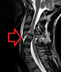
A cervical fracture, commonly called a broken neck, is a fracture of any of the seven cervical vertebrae in the neck. Examples of common causes in humans are traffic collisions and diving into shallow water. Abnormal movement of neck bones or pieces of bone can cause a spinal cord injury, resulting in loss of sensation, paralysis, or usually death soon thereafter, primarily via compromising neurological supply to the respiratory muscles as well as innervation to the heart.

External fixation is a surgical treatment wherein Kirschner pins and wires are inserted and affixed into bone and then exit the body to be attached to an external apparatus composed of rings and threaded rods — the Ilizarov apparatus, the Taylor Spatial Frame, and the Octopod External Fixator — which immobilises the damaged limb to facilitate healing. As an alternative to internal fixation, wherein bone-stabilising mechanical components are surgically emplaced in the body of the patient, external fixation is used to stabilize bone tissues and soft tissues at a distance from the site of the injury.
Advanced trauma life support (ATLS) is a training program for medical providers in the management of acute trauma cases, developed by the American College of Surgeons. Similar programs exist for immediate care providers such as paramedics. The program has been adopted worldwide in over 60 countries, sometimes under the name of Early Management of Severe Trauma, especially outside North America. Its goal is to teach a simplified and standardized approach to trauma patients. Originally designed for emergency situations where only one doctor and one nurse are present, ATLS is now widely accepted as the standard of care for initial assessment and treatment in trauma centers. The premise of the ATLS program is to treat the greatest threat to life first. It also advocates that the lack of a definitive diagnosis and a detailed history should not slow the application of indicated treatment for life-threatening injury, with the most time-critical interventions performed early.

A trauma team is a multidisciplinary group of healthcare workers under the direction of a team leader that works together to assess and treat the severely injured. This team typically meets before the patient reaches the trauma center. Upon arrival, the team does an initial assessment and necessary resuscitation, adhering to a defined protocol.
A traction splint most commonly refers to a splinting device that uses straps attaching over the pelvis or hip as an anchor, a metal rod(s) to mimic normal bone stability and limb length, and a mechanical device to apply traction to the limb.

Blunt trauma, also known as blunt force trauma or non-penetrating trauma, describes a physical trauma due to a forceful impact without penetration of the body's surface. Blunt trauma stands in contrast with penetrating trauma, which occurs when an object pierces the skin, enters body tissue, and creates an open wound. Blunt trauma occurs due to direct physical trauma or impactful force to a body part. Such incidents often occur with road traffic collisions, assaults, and sports-related injuries, and are notably common among the elderly who experience falls.
A pelvic fracture is a break of the bony structure of the pelvis. This includes any break of the sacrum, hip bones, or tailbone. Symptoms include pain, particularly with movement. Complications may include internal bleeding, injury to the bladder, or vaginal trauma.
Focused assessment with sonography in trauma is a rapid bedside ultrasound examination performed by surgeons, emergency physicians, and paramedics as a screening test for blood around the heart or abdominal organs (hemoperitoneum) after trauma. There is also the extended FAST (eFAST) which includes some additional ultrasound views to assess for pneumothorax.
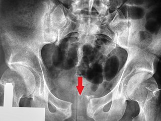
Pubic symphysis diastasis is the separation of normally joined pubic bones, as in the dislocation of the bones, without a fracture that measures radiologically more than 10 mm. Separation of the symphysis pubis is a rare pathology associated with childbirth and has an incidence of 1 in 300 to 1 in 30,000 births. It is usually noticed after delivery but can be observed up to six months postpartum. Risk factors associated with this injury include cephalopelvic disproportion, rapid second stage of labor, epidural anesthesia, severe abduction of the thighs during delivery, or previous trauma to the pelvis. Common signs and symptoms include symphyseal pain aggravated by weight-bearing and walking, a waddling gait, pubic tenderness, and a palpable interpubic gap. Treatment for pubic symphysis diastasis is largely conservative, with treatment modalities including pelvic bracing, bed rest, analgesia, physical therapy, and in some severe cases, surgery.

A tourniquet is a device that is used to apply pressure to a limb or extremity in order to create ischemia or stopping the flow of blood. It may be used in emergencies, in surgery, or in post-operative rehabilitation.

A femoral fracture is a bone fracture that involves the femur. They are typically sustained in high-impact trauma, such as car crashes, due to the large amount of force needed to break the bone. Fractures of the diaphysis, or middle of the femur, are managed differently from those at the head, neck, and trochanter; those are conventionally called hip fractures. Thus, mentions of femoral fracture in medicine usually refer implicitly to femoral fractures at the shaft or distally.

A liver injury, also known as liver laceration, is some form of trauma sustained to the liver. This can occur through either a blunt force such as a car accident, or a penetrating foreign object such as a knife. Liver injuries constitute 5% of all traumas, making it the most common abdominal injury. Generally nonoperative management and observation is all that is required for a full recovery.

Resuscitative endovascular balloon occlusion of the aorta (REBOA) is a minimally invasive procedure performed during resuscitation of critically injured trauma patients. Originally developed as a less invasive alternative to emergency thoracotomy with aortic cross clamping, REBOA is performed to gain rapid control of non-compressible truncal or junctional hemorrhage. REBOA is performed first by achieving access to the common femoral artery (CFA) and advancing a catheter within the aorta. Upon successful catheter placement, an occluding balloon may be inflated either within the descending thoracic aorta or infrarenal abdominal aorta. REBOA stanches downstream hemorrhage and improves cardiac index, cerebral perfusion, and coronary perfusion. Although REBOA does not eliminate the need for definitive hemorrhage control, it may serve as a temporizing measure during initial resuscitation. Despite the benefits of REBOA, there are significant local and systemic ischemic risks. Establishing standardized REBOA procedural indications and mitigating the risk of ischemic injury are topics of ongoing investigation. Although this technique has been successfully deployed in adult patients, it has not yet been studied in children.
References
- 1 2 3 4 5 ATLS - Advanced Trauma Life Support - Student Course Manual (10 ed.). American College of Surgeons. 2018. pp. 96–97. ISBN 9780996826235.
- 1 2 3 4 5 6 7 8 Naseem H, Nesbitt PD, Sprott DC, Clayson A (February 2018). "An assessment of pelvic binder placement at a UK major trauma centre". Annals of the Royal College of Surgeons of England. 100 (2): 101–105. doi:10.1308/rcsann.2017.0159. PMC 5838689 . PMID 29022794.
- 1 2 Walls R, Hockberger R, Gausche-Hill M (2017). Rosen's Emergency Medicine - Concepts and Clinical Practice E-Book. Elsevier Health Sciences. p. 577, 588. ISBN 978-0-323-39016-3.
- ↑ Vaidya R, Roth M, Zarling B, Zhang S, Walsh C, Macsuga J, Swartz J (November 2016). "Application of Circumferential Compression Device (Binder) in Pelvic Injuries: Room for Improvement". The Western Journal of Emergency Medicine. 17 (6): 766–774. doi:10.5811/westjem.2016.7.30057. PMC 5102606 . PMID 27833687.
- ↑ Bonner TJ, Eardley WG, Newell N, Masouros S, Matthews JJ, Gibb I, Clasper JC (November 2011). "Accurate placement of a pelvic binder improves reduction of unstable fractures of the pelvic ring". The Journal of Bone and Joint Surgery. British Volume. 93 (11): 1524–8. doi: 10.1302/0301-620X.93B11.27023 . PMID 22058306.
- ↑ White CE, Hsu JR, Holcomb JB (October 2009). "Haemodynamically unstable pelvic fractures". Injury. 40 (10): 1023–30. doi:10.1016/j.injury.2008.11.023. PMID 19371871.