Related Research Articles

The German Shepherd, also known in Britain as an Alsatian, is a German breed of working dog of medium to large size. The breed was developed by Max von Stephanitz using various traditional German herding dogs from 1899.

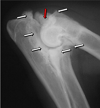
Legg–Calvé–Perthes disease (LCPD) is a childhood hip disorder initiated by a disruption of blood flow to the head of the femur. Due to the lack of blood flow, the bone dies and stops growing. Over time, healing occurs by new blood vessels infiltrating the dead bone and removing the necrotic bone which leads to a loss of bone mass and a weakening of the femoral head.

In dogs, hip dysplasia is an abnormal formation of the hip socket that, in its more severe form, can eventually cause lameness and arthritis of the joints. It is a genetic (polygenic) trait that is affected by environmental factors. It is common in many dog breeds, particularly the larger breeds, and is the most common single cause of arthritis of the hips.

In vertebrate anatomy, the hip, or coxa in medical terminology, refers to either an anatomical region or a joint on the outer (lateral) side of the pelvis.

The health of dogs is a well studied area in veterinary medicine.
Elbow dysplasia is a condition involving multiple developmental abnormalities of the elbow-joint in the dog, specifically the growth of cartilage or the structures surrounding it. These abnormalities, known as 'primary lesions', give rise to osteoarthritic processes. Elbow dysplasia is a common condition of certain breeds of dogs.
Hip scoring is a procedure used to determine the degree of hip dysplasia in dogs and other animals and reporting the findings in a standard way. The hip score is the sum of the points awarded for each of nine radiographic features of both hip joints.
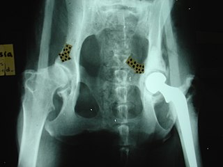
Hip replacement is a surgical procedure performed in dogs and cats as a salvage procedure, to alleviate severe pain in the hip due to, for example, hip dysplasia or irreparable bone fracture. The procedure replaces the head of the femur and the acetabulum with prosthetic implants. Because animals under about 40 pounds (18 kg) carry their own weight with little strain on each leg, hip modification surgeries are often sufficient to restore hip function in many cases. As a result, while hip replacement on animals can be seen in any animal of any size, from cats upwards, it is most often performed in the medium-large breeds of dogs.
Organ replacement in animals is an emerging field in veterinary science, focusing on improving and prolonging the lives of animals through the replacement or augmentation of damaged or dysfunctional organs. Despite its relative rarity compared to limb prosthesis, strides have been made over the decades, with notable milestones such as the first pacemaker surgery on a dog in 1968 and successful kidney transplants in cats since the mid-1980s. This field faces challenges, particularly in canine programs, due to issues related to immunosuppression. Ethical considerations also exist, particularly concerning the treatment of donor animals, underscoring the need for ongoing discussions and regulations in this dynamic field.

Cruciate ligaments are pairs of ligaments arranged like a letter X. They occur in several joints of the body, such as the knee joint, wrist joint and the atlanto-axial joint. In a fashion similar to the cords in a toy Jacob's ladder, the crossed ligaments stabilize the joint while allowing a very large range of motion.

Veterinary surgery is surgery performed on non-human animals by veterinarians, whereby the procedures fall into three broad categories: orthopaedics, soft tissue surgery, and neurosurgery. Advanced surgical procedures such as joint replacement, fracture repair, stabilization of cranial cruciate ligament deficiency, oncologic (cancer) surgery, herniated disc treatment, complicated gastrointestinal or urogenital procedures, kidney transplant, skin grafts, complicated wound management, and minimally invasive procedures are performed by veterinary surgeons. Most general practice veterinarians perform routine surgeries such as neuters and minor mass excisions; some also perform additional procedures.

The Orthopedic Foundation for Animals (OFA) is a nonprofit organization based in Columbia, Missouri, that aims to research and prevent orthopedic and hereditary diseases in companion animals.
A femoral head ostectomy is a surgical operation to remove the head and neck from the femur. It is performed to alleviate pain, and is a salvage procedure, reserved for condition where pain can not be alleviated in any other way. It is common in veterinary surgery. Other names are excision arthroplasty of the femoral head and neck, Girdlestone's operation, Girdlestone procedure, and femoral head and neck ostectomy.
Dislocation of hip may occur in domestic animals.

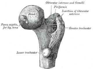
The femoral head is the highest part of the thigh bone (femur). It is supported by the femoral neck.
Protrusio acetabuli is an uncommon defect of the acetabulum, the socket that receives the femoral head to make the hip joint. The hip bone of the pelvic bone/girdle is composed of three bones, the ilium, the ischium and the pubis. In protrusio deformity, there is medial displacement of the femoral head in that the medial aspect of the femoral cortex is medial to the ilioischial line. The socket is too deep and may protrude into the pelvis.

Hip dysplasia is an abnormality of the hip joint where the socket portion does not fully cover the ball portion, resulting in an increased risk for joint dislocation. Hip dysplasia may occur at birth or develop in early life. Regardless, it does not typically produce symptoms in babies less than a year old. Occasionally one leg may be shorter than the other. The left hip is more often affected than the right. Complications without treatment can include arthritis, limping, and low back pain. Females are affected more often than males. Risk factors for hip dysplasia include female sex, family history, certain swaddling practices, and breech presentation whether an infant is delivered vaginally or by cesarean section. If one identical twin is affected, there is a 40% risk the other will also be affected. Screening all babies for the condition by physical examination is recommended. Ultrasonography may also be useful.

Physical therapy for canines adapts human physical therapy techniques to increase function and mobility of joints and muscles in animals. Animal rehabilitation can reduce pain and enhance recovery from injury, surgery, degenerative diseases, age-related diseases, and obesity.

Pain in the hip is the experience of pain in the muscles or joints in the hip/ pelvic region, a condition commonly arising from any of a number of factors. Sometimes it is closely associated with lower back pain.
X-rays of hip dysplasia are one of the two main methods of medical imaging to diagnose hip dysplasia, the other one being medical ultrasonography. Ultrasound imaging yields better results defining the anatomy until the cartilage is ossified. When the infant is around 3 months old a clear roentgenographic image can be achieved. Unfortunately the time the joint gives a good x-ray image is also the point at which nonsurgical treatment methods cease to give good results.
References
- 1 2 3 4 Greer, MA (2014). "PennHIP". Canine Reproduction and Neonatology. CRC Press. p. 24. ISBN 9781498728508.
- ↑ "AIS | PennHIP". antechimagingservices.com. Retrieved December 2, 2019.
- 1 2 Smith, GK (2000). "Hip dysplasia diagnosis - the use of distraction radiography". In Morgan, JP; Wind, A; Davidson, AP (eds.). Hereditary bone and joint diseases in the dog: osteochondroses, hip dysplasia, elbow dysplasia . Hannover: Schlütersche. pp. 300–303. ISBN 3-87706-548-1.
- 1 2 Johnston, Spencer A., editor. Tobias, Karen M., editor. (June 14, 2017). Veterinary surgery : small animal volume one. ISBN 978-0-323-32049-8. OCLC 1030809494.
{{cite book}}:|last=has generic name (help)CS1 maint: multiple names: authors list (link) - ↑ Harasen, Greg (April 2009). "Assessing the dysplastic hip". The Canadian Veterinary Journal. 50 (4): 427–428. PMC 2657530 . PMID 19436454.
- ↑ "PennVet | Pioneering the Diagnosis of Canine Hip Dysplasia". www.vet.upenn.edu. Retrieved December 2, 2019.
- ↑ Sánchez Alonso, C.; García Luque, A.; Chamorro Sancho, M.J.; Arias Sanz, P.; De Vega Terán, P.; Crespo Castejón, F. (September 2015). "El Pennhip modificado y la sinfisiodesis juvenil pubiana como prevención de displasia de cadera canina en las Fuerzas Armadas" [Modified Pennhip and juvenile pubic symphysiodesis as canine hip dysplasia prevention in the Armed Forces]. Sanidad Militar (in Spanish). 71 (3): 146–157. doi: 10.4321/S1887-85712015000300002 .
- 1 2 Harari, Joseph (2000). Small Animal Surgery Secrets. Hanely & Belfus, INC. ISBN 1-56053-355-2.
- ↑ Harasen, Greg (April 2009). "Assessing the dysplastic hip". The Canadian Veterinary Journal. 50 (4): 427–428. PMC 2657530 . PMID 19436454.
- ↑ Williams, Ben (November 14, 2010). "Study compares PennHIP vs OFA hip dysplasia tests". American Animal Hospital Association. Retrieved December 2, 2019.