Peptide loading complex

Last updated
PLC side view PLC side view.jpg
PLC side view

The peptide-loading complex (PLC) is a short-lived, multisubunit membrane protein complex that is located in the endoplasmic reticulum (ER). It orchestrates peptide translocation and selection by major histocompatibility complex class I (MHC-I) molecules. Stable peptide-MHC I complexes are released to the cell surface to promote T-cell response against malignant or infected cells. In turn, T-cells recognize the activated peptides, which could be immunogenic or non-immunogenic.

Contents

Overview

A PLC assembly consists of seven subunits, including the transporters associated with antigen processing (TAP1 and TAP2 – jointly referred to as TAP), the oxidoreductase ERp57, the MHC-I heterodimer, and the chaperones tapasin and calreticulin. TAP transports proteasomal degradation products from the cytosol into the lumen of the ER, where they are loaded onto MHC-I molecules. The peptide-MHC-I complexes then move via a secretory pathway to the cell surface, presenting their antigenic load to cytotoxic T-cells.

In general, preliminary MHC-I heavy chains are chaperoned by the calnexin–calreticulin system in the ER. Together with β2-microglobulin (β2m), MHC-I heavy chains form assemblies of heterodimers that act as receptors for antigenic peptides. Empty MHC-I heterodimers are recruited by calreticulin and form short-lived macromolecular PLC where the chaperone tapasin further provides stabilization in the MHC-I molecules. Furthermore, ERp57 and tapasin form disulfide-linked conjugates, and tapasin is crucial for maintaining the structural stability of the PLC as well as facilitating optimal peptide loading. After final quality control, during which MHC-I heterodimers undergo peptide editing, stable peptide–MHC-I complexes are released to the cell surface for T-cell recognition. The PLC can serve a large variety of MHC-I allomorphs, thus playing a central role in the differentiation and priming of T lymphocytes, and in controlling viral infections and tumour development. [1]

Structure

The structure of the human PLC has been determined using single-particle electron cryo-microscopy (cryo-EM). [2] The PLC, measuring 150 Å by 150 Å and with a total height of 240 Å, is organized around the Transporter associated with Antigen Processing (TAP). It includes molecules such as tapasin, calreticulin, ERp57, and Major Histocompatibility Complex class I (MHC-I), arranged in a pseudo-symmetric pattern.

TAP

TAP is a heterodimeric complex, consisting of TAP1 (ABCB2) and TAP2 (ABCB3) members of the ABC transporter superfamily. The common feature of all ABC transporters is their organization: 1) into two transmembrane domains (TMDs) and 2) into two nucleotide-binding domains (NBDs). Both intramolecular domains are coupled to each other and when ATP binding is in progress, conformational changes in the TMDs allow proteasomal degradation products to move across the membrane. TAP recognizes and transports the antigen peptides produced in the cytosol straight into the ER, while tapasin recognizes the kind of peptides that have the ability to form stable complexes with MHC-I. This process is known as peptide proofreading or editing. Peptides selected through proofreading [3] improve MHC-I stability; tapasin also contributes to the editing of immunogenic peptide epitopes. However, only lately it was proven via biochemical, biophysical, and structural studies that a key function in adaptive immunity, the catalytic mechanism of peptide proofreading, is performed by tapasin and TAPBPR (TAP-binding protein-related, a tapasin homologue). [4]

Tapasin

Cresswell and co-workers first discovered tapasin (TAP-associated glycoprotein) as a 48 kDa protein in complexes isolated with TAP1 antibodies from digitonin lysates of human B lymphoblastoid cells. [5] Tapasin binds HC/β2m along with ER chaperones to the peptide transporter. [6] It is located in the ER and its function comprises holding together class I molecules jointly with the chaperone calreticulin and the ERp57 to TAP. Studies of a tapasin-deficient cell line and from mice bearing a disrupted tapasin gene, the short-lived complex of class I molecules.[ clarification needed ]

Tapasin and TAP are very important for the stabilization of the class I molecules and also for the optimization of the peptide presented to cytotoxic T cells. [7] A PLC-independent tapasin homologue protein named TAPBPR [4] was found that has the ability to act as a second MHC-I specific peptide proofreader or editor, but does not possess a transmembrane domain. [8] Tapasin and TAPBPR [4] share similar binding interfaces on MHC-I, as shown with the X-ray structure of TAPBPR with MHC-I (heavy chain and β2 microglobulin). The use of a photo-cleavable high-affinity peptide allowed researchers to form a stable (bound) MHC-I molecules and afterwards to form a stable TAPBPR [4] and MHC-I complex with cleavage by UV light of the photoinduced peptide.

ERp57

Crystal structure of Tapasin-ERp57 complex Tapasin-ERp57 structure.png
Crystal structure of Tapasin-ERp57 complex

ERp57 is an enzyme of the thiol oxidoreductase family located in the ER. [9] It is attached to substrates in an indirect fashion through association with the molecular chaperone calreticulin of the peptide-loading complex, [10] [11] In early stages of generation of MHC-I molecules, ERp57 is associated with free MHC-I heavy chains. As a result, its function is determined by the formation of disulfide bonds in heavy chains, by oxidative folding of the heavy chain, and finally by the fact that ERp57 is loading the peptides onto MHC-I molecules.

MHC-I

MHC-I heavy chains may work as chaperones with the aid of the calnexin-calreticulin complex in the ER. In addition to this, β2-microglobulin (β2m) is attached to the heavy chains of the heterodimers and as a whole they act as receptors for antigenic peptides. When MHC-I chains are empty, they are recruited by calreticulin and form a transient PLC.

Release of MHC-I from PLC PLC schematics.png
Release of MHC-I from PLC

Tapasin regularly plays a role in the stabilization of MHC-I. Only after MHC-I heterodimers are deployed for peptide proofreading or editing, stable pMHC-I (peptide-MHC-I) complexes are released to the cell surface for recognition and destruction of virus-infected or malignantly neoplastic cells. In general, each individual organism owns a collection of six MHC-I molecules (three from each parent). Thus, in autoimmune emergencies, compatible donors are relatives who own a similar collection of MHC-I molecules, apart from those of the recipient.[ citation needed ]

Calreticulin

Calreticulin – especially its lectin-like domain – interacts with MHC-I. The P domain faces the MHC-I peptide-binding site towards ERp57. This orientation makes it possible for tapasin to attach and secure MHC-I. This translocation of TAP facilitates its opening out into an ER luminal cavity, edged by standard membrane entry points such as those for tapasin and MHC-I. These two entry points facilitate the recruitment of MHC-I with optimal peptide loading and eventual release of MHC-I in T-cell surfaces for recognition.[ citation needed ]

Related Research Articles

<span class="mw-page-title-main">Calreticulin</span> Soluble protein

Calreticulin also known as calregulin, CRP55, CaBP3, calsequestrin-like protein, and endoplasmic reticulum resident protein 60 (ERp60) is a protein that in humans is encoded by the CALR gene.

<span class="mw-page-title-main">Major histocompatibility complex</span> Cell surface proteins, part of the acquired immune system

The major histocompatibility complex (MHC) is a large locus on vertebrate DNA containing a set of closely linked polymorphic genes that code for cell surface proteins essential for the adaptive immune system. These cell surface proteins are called MHC molecules.

Antigen processing, or the cytosolic pathway, is an immunological process that prepares antigens for presentation to special cells of the immune system called T lymphocytes. It is considered to be a stage of antigen presentation pathways. This process involves two distinct pathways for processing of antigens from an organism's own (self) proteins or intracellular pathogens, or from phagocytosed pathogens ; subsequent presentation of these antigens on class I or class II major histocompatibility complex (MHC) molecules is dependent on which pathway is used. Both MHC class I and II are required to bind antigens before they are stably expressed on a cell surface. MHC I antigen presentation typically involves the endogenous pathway of antigen processing, and MHC II antigen presentation involves the exogenous pathway of antigen processing. Cross-presentation involves parts of the exogenous and the endogenous pathways but ultimately involves the latter portion of the endogenous pathway.

<span class="mw-page-title-main">MHC class I</span> Protein of the immune system

MHC class I molecules are one of two primary classes of major histocompatibility complex (MHC) molecules and are found on the cell surface of all nucleated cells in the bodies of vertebrates. They also occur on platelets, but not on red blood cells. Their function is to display peptide fragments of proteins from within the cell to cytotoxic T cells; this will trigger an immediate response from the immune system against a particular non-self antigen displayed with the help of an MHC class I protein. Because MHC class I molecules present peptides derived from cytosolic proteins, the pathway of MHC class I presentation is often called cytosolic or endogenous pathway.

<span class="mw-page-title-main">T-cell receptor</span> Protein complex on the surface of T cells that recognizes antigens

The T-cell receptor (TCR) is a protein complex found on the surface of T cells, or T lymphocytes, that is responsible for recognizing fragments of antigen as peptides bound to major histocompatibility complex (MHC) molecules. The binding between TCR and antigen peptides is of relatively low affinity and is degenerate: that is, many TCRs recognize the same antigen peptide and many antigen peptides are recognized by the same TCR.

Cross-presentation is the ability of certain professional antigen-presenting cells (mostly dendritic cells) to take up, process and present extracellular antigens with MHC class I molecules to CD8 T cells (cytotoxic T cells). Cross-priming, the result of this process, describes the stimulation of naive cytotoxic CD8+ T cells into activated cytotoxic CD8+ T cells. This process is necessary for immunity against most tumors and against viruses that infect dendritic cells and sabotage their presentation of virus antigens. Cross presentation is also required for the induction of cytotoxic immunity by vaccination with protein antigens, for example, tumour vaccination.

<span class="mw-page-title-main">Calnexin</span> Mammalian protein found in Homo sapiens

Calnexin (CNX) is a 67kDa integral protein of the endoplasmic reticulum (ER). It consists of a large N-terminal calcium-binding lumenal domain, a single transmembrane helix and a short, acidic cytoplasmic tail. In humans, calnexin is encoded by the gene CANX.

<span class="mw-page-title-main">Antigen presentation</span> Vital immune process that is essential for T cell immune response triggering

Antigen presentation is a vital immune process that is essential for T cell immune response triggering. Because T cells recognize only fragmented antigens displayed on cell surfaces, antigen processing must occur before the antigen fragment can be recognized by a T-cell receptor. Specifically, the fragment, bound to the major histocompatibility complex (MHC), is transported to the surface of the cell Antigen-Presenting Cell, a process known as presentation. If there has been an infection with viruses or bacteria, the cell Antigen-Presenting Cell will present an endogenous or exogenous peptide fragment derived from the antigen by MHC molecules. There are two types of MHC molecules which differ in the behaviour of the antigens: MHC class I molecules (MHC-I) bind peptides from the cell cytosol, while peptides generated in the endocytic vesicles after internalisation are bound to MHC class II (MHC-II). Cellular membranes separate these two cellular environments - intracellular and extracellular. Each T cell can only recognize tens to hundreds of copies of a unique sequence of a single peptide among thousands of other peptides presented on the same cell, because an MHC molecule in one cell can bind to quite a large range of peptides. Predicting which antigens will be presented to the immune system by a certain MHC/HLA type is difficult, but the technology involved is improving.

<span class="mw-page-title-main">MHC class II</span> Protein of the immune system

MHC Class II molecules are a class of major histocompatibility complex (MHC) molecules normally found only on professional antigen-presenting cells such as dendritic cells, macrophages, some endothelial cells, thymic epithelial cells, and B cells. These cells are important in initiating immune responses.

Transporter associated with antigen processing (TAP) protein complex belongs to the ATP-binding-cassette transporter family. It delivers cytosolic peptides into the endoplasmic reticulum (ER), where they bind to nascent MHC class I molecules.

<span class="mw-page-title-main">HLA-A</span> Protein-coding gene in the species Homo sapiens

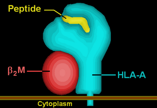
HLA-A is a group of human leukocyte antigens (HLA) that are encoded by the HLA-A locus, which is located at human chromosome 6p21.3. HLA is a major histocompatibility complex (MHC) antigen specific to humans. HLA-A is one of three major types of human MHC class I transmembrane proteins. The others are HLA-B and HLA-C. The protein is a heterodimer, and is composed of a heavy α chain and smaller β chain. The α chain is encoded by a variant HLA-A gene, and the β chain (β2-microglobulin) is an invariant β2 microglobulin molecule. The β2 microglobulin protein is encoded by the B2M gene, which is located at chromosome 15q21.1 in humans.

<span class="mw-page-title-main">CLIP (protein)</span>

CLIP or Class II-associated invariant chain peptide is the part of the invariant chain (Ii) that binds to the peptide binding groove of MHC class II and remains there until the MHC receptor is fully assembled. CLIP is one of the most prevalent self peptides found in the thymic cortex of most antigen-presenting cells. The purpose of CLIP is to prevent the degradation of MHC II dimers before antigenic peptides bind, and to prevent autoimmunity.

<span class="mw-page-title-main">HLA-E</span> Protein-coding gene in the species Homo sapiens

HLA class I histocompatibility antigen, alpha chain E (HLA-E) also known as MHC class I antigen E is a protein that in humans is encoded by the HLA-E gene. The human HLA-E is a non-classical MHC class I molecule that is characterized by a limited polymorphism and a lower cell surface expression than its classical paralogues. The functional homolog in mice is called Qa-1b, officially known as H2-T23.

<span class="mw-page-title-main">Tapasin</span> Type of protein

TAP-associated glycoprotein, also known as tapasin or TAPBP, is a protein that in humans is encoded by the TAPBP gene.

<span class="mw-page-title-main">PDIA3</span> Protein-coding gene in the species Homo sapiens

Protein disulfide-isomerase A3 (PDIA3), also known as glucose-regulated protein, 58-kD (GRP58), is an isomerase enzyme encoded by the autosomal gene PDIA3 in humans. This protein localizes to the endoplasmic reticulum (ER) and interacts with lectin chaperones calreticulin and calnexin (CNX) to modulate folding of newly synthesized glycoproteins. It is thought that complexes of lectins and this protein mediate protein folding by promoting formation of disulfide bonds in their glycoprotein substrates.

<span class="mw-page-title-main">HLA-DM</span>

HLA-DM is an intracellular protein involved in the mechanism of antigen presentation on antigen presenting cells (APCs) of the immune system. It does this by assisting in peptide loading of major histocompatibility complex (MHC) class II membrane-bound proteins. HLA-DM is encoded by the genes HLA-DMA and HLA-DMB.

<span class="mw-page-title-main">TAP1</span> Protein-coding gene in the species Homo sapiens

Transporter associated with antigen processing 1 (TAP1) is a protein that in humans is encoded by the TAP1 gene. A member of the ATP-binding cassette transporter family, it is also known as ABCB2.

<span class="mw-page-title-main">HLA-F</span> Protein-coding gene in the species Homo sapiens

HLA class I histocompatibility antigen, alpha chain F is a protein that in humans is encoded by the HLA-F gene. It is an empty intracellular molecule that encodes a non-classical heavy chain anchored to the membrane and forming a heterodimer with a β-2 microglobulin light chain. It belongs to the HLA class I heavy chain paralogues that separate from most of the HLA heavy chains. HLA-F is localized in the endoplasmic reticulum and Golgi apparatus, and is also unique in the sense that it exhibits few polymorphisms in the human population relative to the other HLA genes; however, there have been found different isoforms from numerous transcript variants found for the HLA-F gene. Its pathways include IFN-gamma signaling and CDK-mediated phosphorylation and removal of the Saccharomycescerevisiae Cdc6 protein, which is crucial for functional DNA replication.

<span class="mw-page-title-main">TAP2</span> Protein-coding gene in the species Homo sapiens

TAP2 is a gene in humans that encodes the protein Antigen peptide transporter 2.

Immunoevasins are proteins expressed by some viruses that enable the virus to evade immune recognition by interfering with MHC I complexes in the infected cell, therefore blocking the recognition of viral protein fragments by CD8+ cytotoxic T lymphocytes. Less frequently, MHC II antigen presentation and induced-self molecules may also be targeted. Some viral immunoevasins block peptide entry into the endoplasmic reticulum (ER) by targeting the TAP transporters. Immunoevasins are particularly abundant in viruses that are capable of establishing long-term infections of the host, such as herpesviruses.

References

  1. Blees, A.; Januliene, D.; Hofmann, T.; Koller, N.; Blanchet, C.; Rupp, B.; Ellenrieder, L.; Dalpke, A.; Zimmermann, R.; Heck, A.; Hillenbrand, M.; Diederichs, K.; Than, M. (2017). "The MHC I peptide loading complex: a multitasking machinery in adaptive immunity". Trends in Biochemical Sciences. 42 (8): 648–665. doi: 10.1016/j.tibs.2017.05.005 .
  2. Blees A, Januliene D, Hofmann T, Koller N, Schmidt C, Trowitzsch S, Moeller A, Tampé R (November 2017). "Structure of the human MHC-I peptide-loading complex". Nature. 551 (7681): 525–528. Bibcode:2017Natur.551..525B. doi:10.1038/nature24627. PMID   29107940. S2CID   4447406.
  3. Thomas C, Tampé R (2017). "Proofreading of Peptide-MHC Complexes through Dynamic Multivalent Interactions". Frontiers in Immunology. 8: 65. doi: 10.3389/fimmu.2017.00065 . PMC   5296336 . PMID   28228754.
  4. 1 2 3 4 Thomas C, Tampé R (November 2017). "Structure of the TAPBPR-MHC I complex defines the mechanism of peptide loading and editing". Science. 358 (6366): 1060–1064. Bibcode:2017Sci...358.1060T. doi: 10.1126/science.aao6001 . PMID   29025996.
  5. Ortmann B, Androlewicz MJ, Cresswell P (April 1994). "MHC class I/beta 2-microglobulin complexes associate with TAP transporters before peptide binding". Nature. 368 (6474): 864–7. Bibcode:1994Natur.368..864O. doi:10.1038/368864a0. PMID   8159247. S2CID   4268516.
  6. Sadasivan B, Lehner PJ, Ortmann B, Spies T, Cresswell P (August 1996). "Roles for calreticulin and a novel glycoprotein, tapasin, in the interaction of MHC class I molecules with TAP". Immunity. 5 (2): 103–14. doi: 10.1016/S1074-7613(00)80487-2 . PMID   8769474.
  7. Momburg F, Tan P (October 2002). "Tapasin-the keystone of the loading complex optimizing peptide binding by MHC class I molecules in the endoplasmic reticulum". Molecular Immunology. 39 (3–4): 217–33. doi:10.1016/S0161-5890(02)00103-7. PMID   12200052.
  8. McShan AC, Natarajan K, Kumirov VK, Flores-Solis D, Jiang J, Badstübner M, et al. (August 2018). "Peptide exchange on MHC-I by TAPBPR is driven by a negative allostery release cycle". Nature Chemical Biology. 14 (8): 811–820. doi:10.1038/s41589-018-0096-2. PMC   6202177 . PMID   29988068.
  9. Frickel EM, Frei P, Bouvier M, Stafford WF, Helenius A, Glockshuber R, Ellgaard L (April 2004). "ERp57 is a multifunctional thiol-disulfide oxidoreductase". The Journal of Biological Chemistry. 279 (18): 18277–87. doi: 10.1074/jbc.M314089200 . PMID   14871896.
  10. Oliver JD, Roderick HL, Llewellyn DH, High S (August 1999). "ERp57 functions as a subunit of specific complexes formed with the ER lectins calreticulin and calnexin". Molecular Biology of the Cell. 10 (8): 2573–82. doi:10.1091/mbc.10.8.2573. PMC   25489 . PMID   10436013.
  11. Zhang Y, Baig E, Williams DB (May 2006). "Functions of ERp57 in the folding and assembly of major histocompatibility complex class I molecules". The Journal of Biological Chemistry. 281 (21): 14622–31. doi: 10.1074/jbc.M512073200 . PMID   16567808.