Related Research Articles

Radiation therapy or radiotherapy, often abbreviated RT, RTx, or XRT, is a therapy using ionizing radiation, generally provided as part of cancer treatment to control or kill malignant cells and normally delivered by a linear accelerator. Radiation therapy may be curative in a number of types of cancer if they are localized to one area of the body. It may also be used as part of adjuvant therapy, to prevent tumor recurrence after surgery to remove a primary malignant tumor. Radiation therapy is synergistic with chemotherapy, and has been used before, during, and after chemotherapy in susceptible cancers. The subspecialty of oncology concerned with radiotherapy is called radiation oncology. A physician who practices in this subspecialty is a radiation oncologist.
In physics, attenuation is the gradual loss of flux intensity through a medium. For instance, dark glasses attenuate sunlight, lead attenuates X-rays, and water and air attenuate both light and sound at variable attenuation rates.
External beam radiation therapy (EBRT) is a compound word that refers to the use of a collimated beam of ionizing radiation from outside the body to treat a disease.
Radiation dosimetry in the fields of health physics and radiation protection is the measurement, calculation and assessment of the ionizing radiation dose absorbed by an object, usually the human body. This applies both internally, due to ingested or inhaled radioactive substances, or externally due to irradiation by sources of radiation.
The therapeutic index is a quantitative measurement of the relative safety of a drug. It is a comparison of the amount of a therapeutic agent that causes the therapeutic effect to the amount that causes toxicity. The related terms therapeutic window or safety window refer to a range of doses optimized between efficacy and toxicity, achieving the greatest therapeutic benefit without resulting in unacceptable side-effects or toxicity.

Radiosurgery is surgery using radiation, that is, the destruction of precisely selected areas of tissue using ionizing radiation rather than excision with a blade. Like other forms of radiation therapy, it is usually used to treat cancer. Radiosurgery was originally defined by the Swedish neurosurgeon Lars Leksell as "a single high dose fraction of radiation, stereotactically directed to an intracranial region of interest".
A monitor unit (MU) is a measure of machine output from a clinical accelerator for radiation therapy such as a linear accelerator or an orthovoltage unit. Monitor units are measured by monitor chambers, which are ionization chambers that measure the dose delivered by a beam and are built into the treatment head of radiotherapy linear accelerators.

A radiation burn is a damage to the skin or other biological tissue and organs as an effect of radiation. The radiation types of greatest concern are thermal radiation, radio frequency energy, ultraviolet light and ionizing radiation.

The Bragg peak is a pronounced peak on the Bragg curve which plots the energy loss of ionizing radiation during its travel through matter. For protons, α-rays, and other ion rays, the peak occurs immediately before the particles come to rest. It is named after William Henry Bragg, who discovered it in 1903.
In passing through matter, charged particles ionize and thus lose energy in many steps, until their energy is (almost) zero. The distance to this point is called the range of the particle. The range depends on the type of particle, on its initial energy and on the material through which it passes.
A point source is a single identifiable localised source of something. A point source has negligible extent, distinguishing it from other source geometries. Sources are called point sources because in mathematical modeling, these sources can usually be approximated as a mathematical point to simplify analysis.

In external beam Radiotherapy, transverse and longitudinal dose measurements are taken by a radiation detector in order to characterise the radiation beams from medical linear accelerators. Typically, an ionisation chamber and water phantom are used to create these radiation dose profiles. Water is used due to its tissue equivalence.

Fast neutron therapy utilizes high energy neutrons typically between 50 and 70 MeV to treat cancer. Most fast neutron therapy beams are produced by reactors, cyclotrons (d+Be) and linear accelerators. Neutron therapy is currently available in Germany, Russia, South Africa and the United States. In the United States, one treatment center is operational, in Seattle, Washington. The Seattle center uses a cyclotron which produces a proton beam impinging upon a beryllium target.

In radiotherapy, radiation treatment planning (RTP) is the process in which a team consisting of radiation oncologists, radiation therapist, medical physicists and medical dosimetrists plan the appropriate external beam radiotherapy or internal brachytherapy treatment technique for a patient with cancer.
Particle therapy is a form of external beam radiotherapy using beams of energetic neutrons, protons, or other heavier positive ions for cancer treatment. The most common type of particle therapy as of August 2021 is proton therapy.
A dose-volume histogram (DVH) is a histogram relating radiation dose to tissue volume in radiation therapy planning. DVHs are most commonly used as a plan evaluation tool and to compare doses from different plans or to structures. DVHs were introduced by Michael Goitein and Verhey in 1979. DVH summarizes 3D dose distributions in a graphical 2D format. In modern radiation therapy, 3D dose distributions are typically created in a computerized treatment planning system (TPS) based on a 3D reconstruction of a CT scan. The "volume" referred to in DVH analysis is a target of radiation treatment, a healthy organ nearby a target, or an arbitrary structure.
Tissue-to-air ratio (TAR) is a term used in radiotherapy treatment planning to help calculate absorbed dose to water in conditions other than those directly measured.
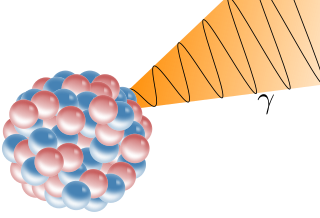
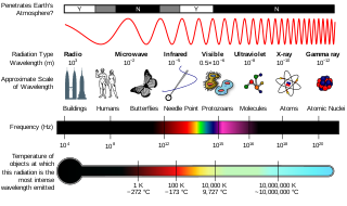
A gamma ray, also known as gamma radiation (symbol γ or ), is a penetrating form of electromagnetic radiation arising from the radioactive decay of atomic nuclei. It consists of the shortest wavelength electromagnetic waves, typically shorter than those of X-rays. With frequencies above 30 exahertz (3×10¹⁹ Hz), it imparts the highest photon energy. Paul Villard, a French chemist and physicist, discovered gamma radiation in 1900 while studying radiation emitted by radium. In 1903, Ernest Rutherford named this radiation gamma rays based on their relatively strong penetration of matter; in 1900 he had already named two less penetrating types of decay radiation (discovered by Henri Becquerel) alpha rays and beta rays in ascending order of penetrating power.
Dose area product (DAP) is a quantity used in assessing the radiation risk from diagnostic X-ray examinations and interventional procedures. It is defined as the absorbed dose multiplied by the area irradiated, expressed in gray-centimetres squared. Manufacturers of DAP meters usually calibrate them in terms of absorbed dose to air. DAP reflects not only the dose within the radiation field but also the area of tissue irradiated. Therefore, it may be a better indicator of the overall risk of inducing cancer than the dose within the field. It also has the advantages of being easily measured, with the permanent installation of a DAP meter on the X-ray set. Due to the divergence of a beam emitted from a "point source", the area irradiated (A) increases with the square of distance from the source, while radiation intensity (I) decreases according to the inverse square of distance. Consequently, the product of intensity and area, and therefore DAP, is independent of distance from the source.
Radiation exposure is a measure of the ionization of air due to ionizing radiation from photons. It is defined as the electric charge freed by such radiation in a specified volume of air divided by the mass of that air. As of 2007, "medical radiation exposure" was defined by the International Commission on Radiological Protection as exposure incurred by people as part of their own medical or dental diagnosis or treatment; by persons, other than those occupationally exposed, knowingly, while voluntarily helping in the support and comfort of patients; and by volunteers in a programme of biomedical research involving their exposure. Common medical tests and treatments involving radiation include X-rays, CT scans, mammography, lung ventilation and perfusion scans, bone scans, cardiac perfusion scan, angiography, radiation therapy, and more. Each type of test carries its own amount of radiation exposure. There are two general categories of adverse health effects caused by radiation exposure: deterministic effects and stochastic effects. Deterministic effects are due to the killing/malfunction of cells following high doses; and stochastic effects involve either cancer development in exposed individuals caused by mutation of somatic cells, or heritable disease in their offspring from mutation of reproductive (germ) cells.
References
- ↑ McDermott, Patrick; Orton, Colin G. (2018). The physics & technology of radiation therapy (Second ed.). p. 10-7. ISBN 978-1930524989.
- [1] Radiation Therapy Physics, Hendee W., Ibbott G. and Hendee E. (2005) Wiley-Liss Publ., ISBN 0-471-39493-9